ffn_cellsim

A fine-grained, mechanistic, GPU-native simulator of single-cell mechanobiology — every filament, motor head, adhesion clutch and cell–cell junction is an explicit particle or bond, not a lumped model.

Mechanistic hard rule. For every design decision, pick the full-fidelity, fine-grained, mechanistic option (explicit particles and bonds, force-dependent kinetics) over any abstracted, lumped, or proxy mechanism — even at higher implementation cost — so that published closed-form models serve only as validation oracles and never as the runtime mechanism.
2GPU-native physics engines · DCM + FF
46explicit mechanisms · no lumped models
18validation checkpoints vs oracles & MCF7
6/6ECM materials validated in real Pascals
266kparticles in the native MCF7 cell
1,400+commits over 11 weeks
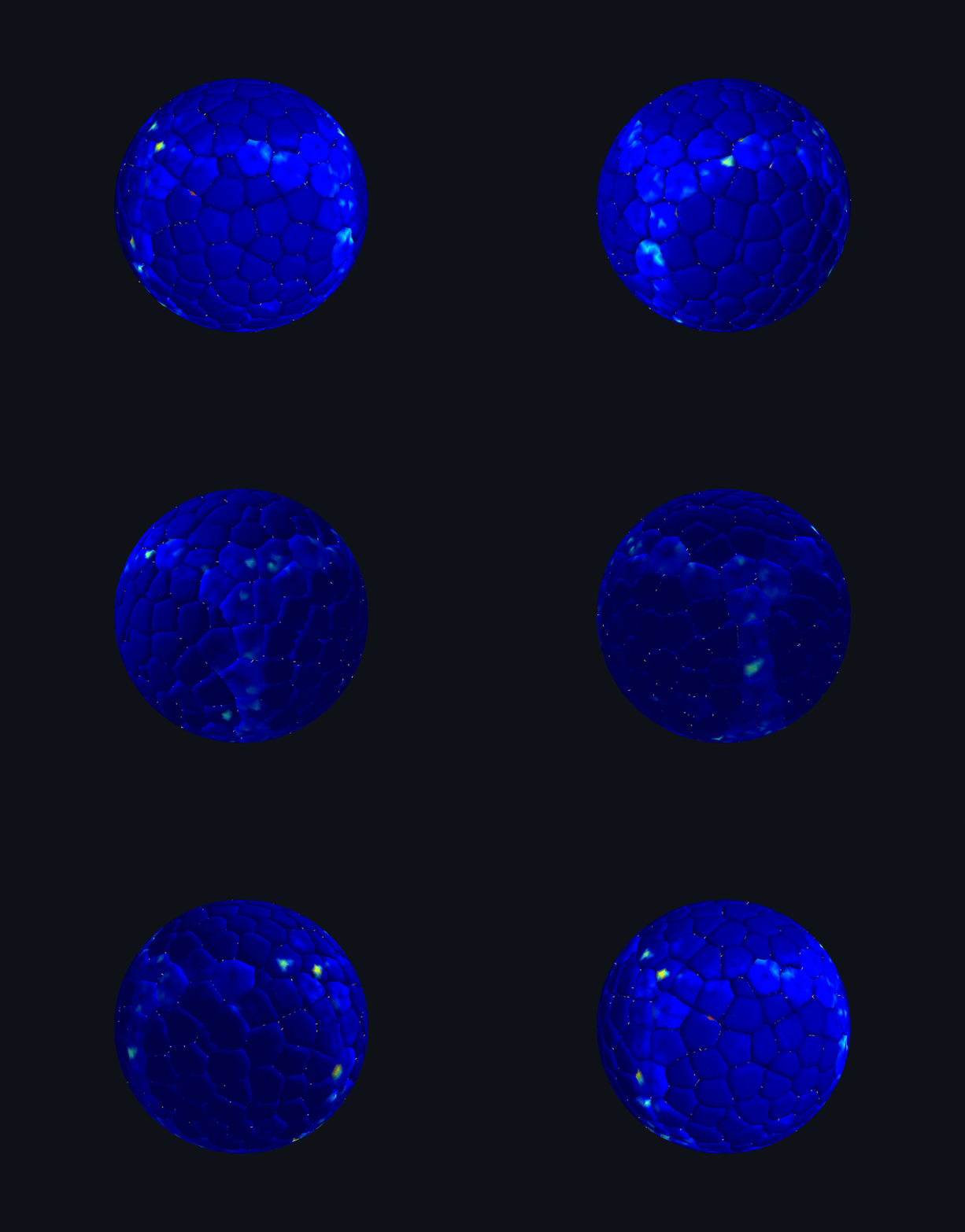
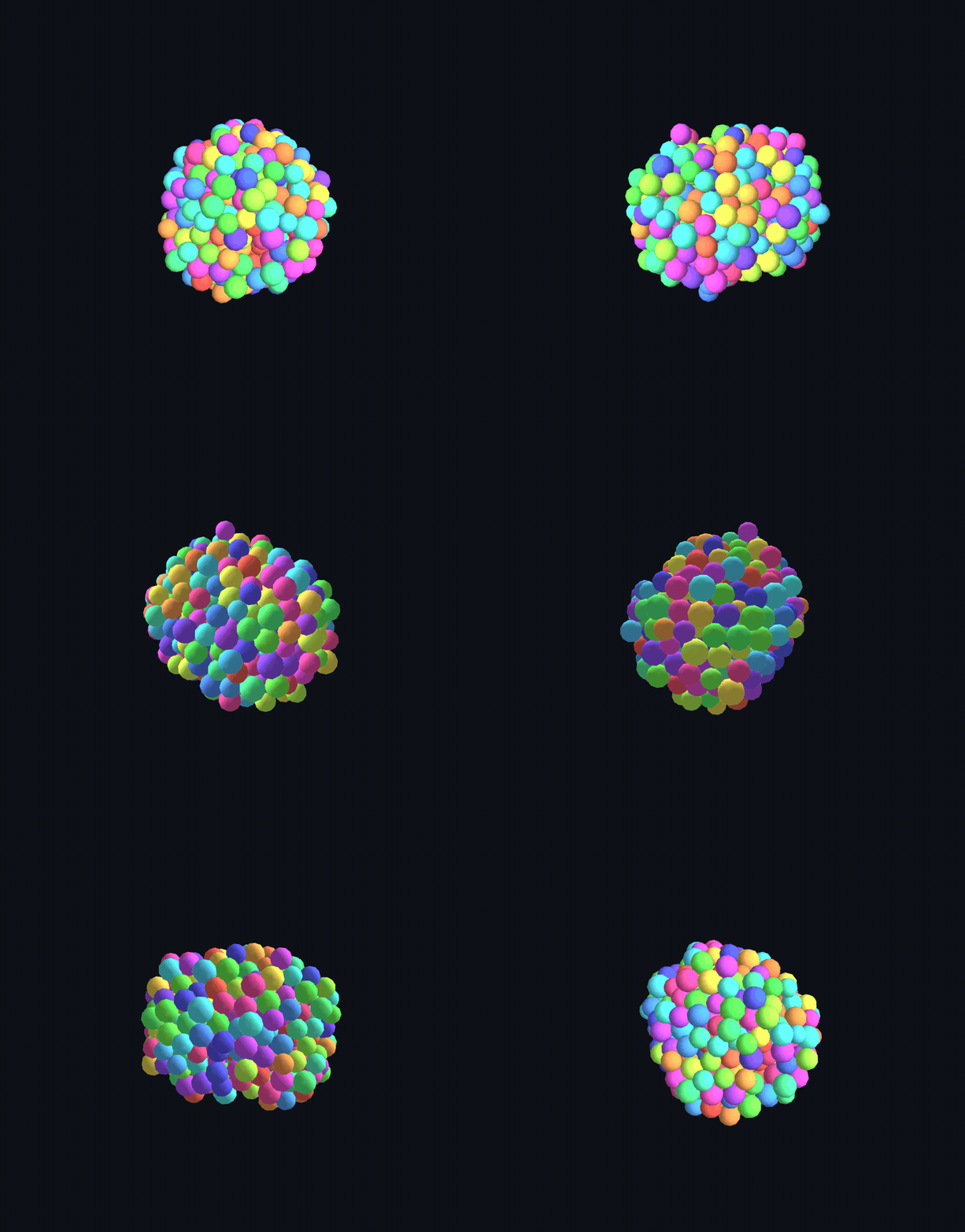
DCM N=2000 confluent spheroid turntable
DCM — N=2,000 confluent spheroid, emergent junction stress
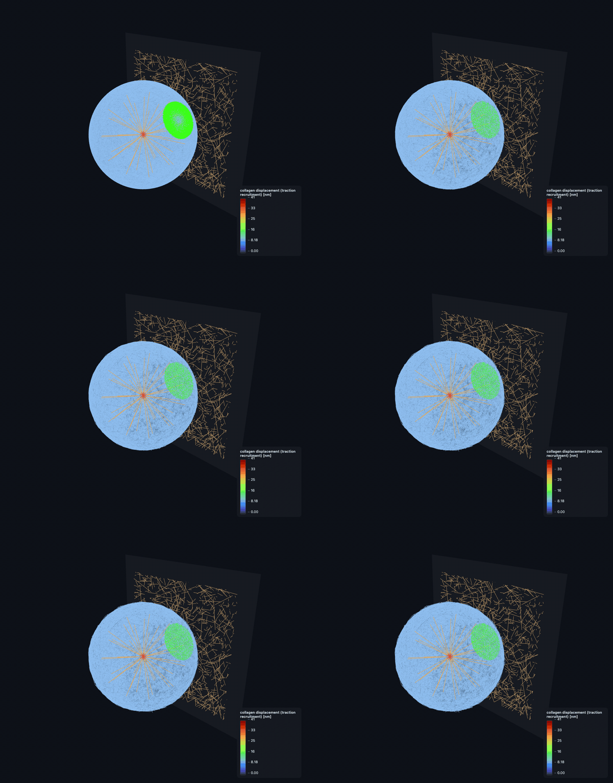
FF cell remodeling a collagen-I matrix
FF — native MCF7 cell recruits & remodels a collagen ECM
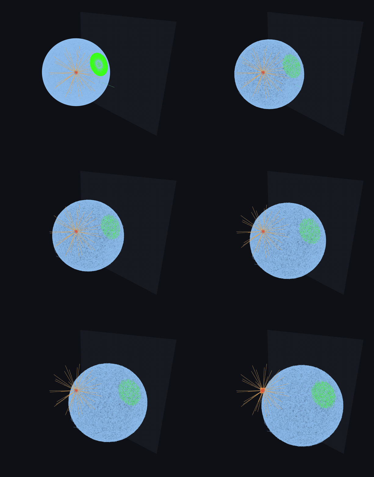
FF cell crawling on a substrate
FF — whole-cell crawl emerges from protrusion + traction

Two GPU-resident physics engines

One continuum prototype (ActiveCellSim v1) became two independent, differentiable, NVIDIA-Warp engines — validated kernel-by-kernel against analytic ground truth and published literature.

DCM — Deformable Cell Model engine (ffn_sim/dcm/)

Physics lineage: Re-implements the deformable-cell-model literature, used as reference only (never imported at runtime): primary physics basis is SimuCell3D (Runser, Vetter & Iber 2024, Nat. Comput. Sci. 4:299-309) — node-face contact, surface-tension faceting (gamma-tilde = gamma/(K*l)), compressible-fluid cytoplasm p = -K*ln(V/V0), measured-value defaults; with CellSim3D (Madhikar, Astrom, Westerholm & Karttunen 2018, Comput. Phys. Commun. 232:206-213) as a cross-check and GPU/division/fixed-topology implementation reference for the Warp port.

Models: The cell as a pressurised elastic shell (NOT resolved filaments): membrane/cortex edge springs, cytoplasmic turgor pressure (with physiological viscosity), nucleus, node-node cohesion and node-face contact between cells, and surface-tension faceting. Scales to many-cell spheroids (~10^2-10^3 deformable cells, each ~42 nodes) on a single RTX A5000. GPU-resident and differentiable on NVIDIA Warp, with a host-side node-pool remesh; every force and integrator kernel is parity-gated against the archived HOOMD-blue reference.

Purpose: The mesoscale, whole-cell-shell / multicellular engine. Scope: emergent aggregation, compaction, faceting, division, and substrate spreading of deformable cells and spheroids, plus differentiable parameter estimation / inverse design. It deliberately does not resolve the cytoskeleton — that is the FF layer's job.

FF — Filament-FEM engine (ffn_sim/ff/)

Physics lineage: Re-implements Cytosim filament physics (Nedelec & Foethke 2007, New J. Phys.), used as reference only (never imported at runtime), as the going-forward fine-grained engine — the mechanistic counterpart to the DCM shell model.

Models: The explicit, fine-grained cytoskeleton: individual actin filaments (bending/stretching mechanics, barbed-end growth), motor heads, cross-linkers, the actin cortex, and the surrounding ECM fiber network, with turgor and Hand-style kinetic Monte-Carlo binding. Filament architectures are woven from shared primitives (filaments + nucleator + crosslinker + motor) into cortex, filopodium (bundled), and lamellipodium (Arp2/3 dendritic branching) forms. GPU-native on NVIDIA Warp, validated at native resolution on an RTX A5000.

Purpose: The fine-grained subcellular engine. Scope: filament-resolved mechanics — cortical tension generation, filopodial/lamellipodial protrusion, and ECM remodeling — where macroscopic quantities (e.g. cortical tension, protrusion force) are meant to emerge from the explicit filament/motor/cross-link dynamics. Build in progress.

Ten weeks · three architectural eras

Era I · Continuum v1
W01

Continuum spheroid platform

Taichi MLS-MPM spheroid on a substrate; 80 h spreading run to 5,000 cells, layered osmotic/ECM chemistry.

Era I · Continuum v1
W02

Validation against experiment

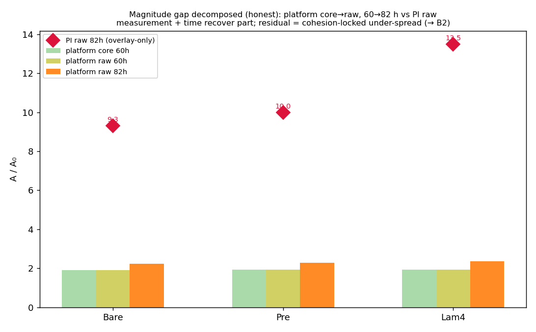
Measurement-matched A/A₀ vs the PI's MCF7 spheroids — surfaced the ~5–9× under-spread ceiling that forced the rebuild.

Era I → II
W03

The mechanistic pivot

Hard rule ratified: replace every lumped/paper-model mechanism with explicit particle/bond physics; first oracle tests land.

Era II · HOOMD
W04

Fine-grained foundations

HOOMD-blue + custom BAOAB integrator: semiflexible filaments, cortex topology, focal-adhesion / motor-clutch (Hill, Bell–Evans).

Era II · HOOMD
W05

AFINES motors, native cortex

AFINES grip-walk; membrane/nucleus/cytoplasm compartments; connected spanning-mesh cortex to ~38k filaments; 44× CUDA integrator.

Era II · HOOMD
W06

Full compartment stack

8 subcellular systems EXPERIMENTAL→LIVE on one MCF7 cell (~25k particles); Gate-B quantifies emergent tension + the myosin γ-floor.

Era III · Warp
W07

GPU-native deformable cells

Ground-up DCM (SimuCell3D) on NVIDIA Warp; node-face cadherin cohesion; two-stage aggregation→spreading recovers A/A₀=a+b/R+c/R².

Era III · Warp
W08

Contact mechanics + ECM

Full IPC (log-barrier + CCD, no interpenetration); single cell coupled to a fiber ECM (Taeyoon-Kim), recovering ~1/r strain decay.

Era III · Warp
W09

GPU-native filaments, faceting

FF (Cytosim) goes fully GPU-native; confluent Voronoi init resolves faceting; proliferation N=400→477, volume conserved.

Era III · Warp
W10

Full-compartment, emergent stress

Both engines mature: per-cell compartment spheroids to N=2,000 with virial von-Mises stress at cadherin junctions; FF filopodium into collagen; AFM full-compartment indentation.

What the framework demonstrates

Four headline results — each an emergent behaviour read out from the explicit particle/bond dynamics, shown with its evidence, including where the model is honestly short of reality.

Emergent cortical tension — and an honest γ-floor

Passive osmotic turgor reproduces the MCF7 cortical-tension band (Young–Laplace γ=ΔP·R/2), but the myosin-active channel comes out ~530× short at the literature prestress — the γ-floor is surfaced and documented, never tuned away.

See the validation ↓

A cell recruits & remodels its own collagen ECM

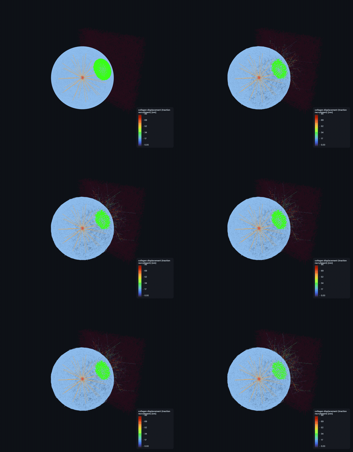
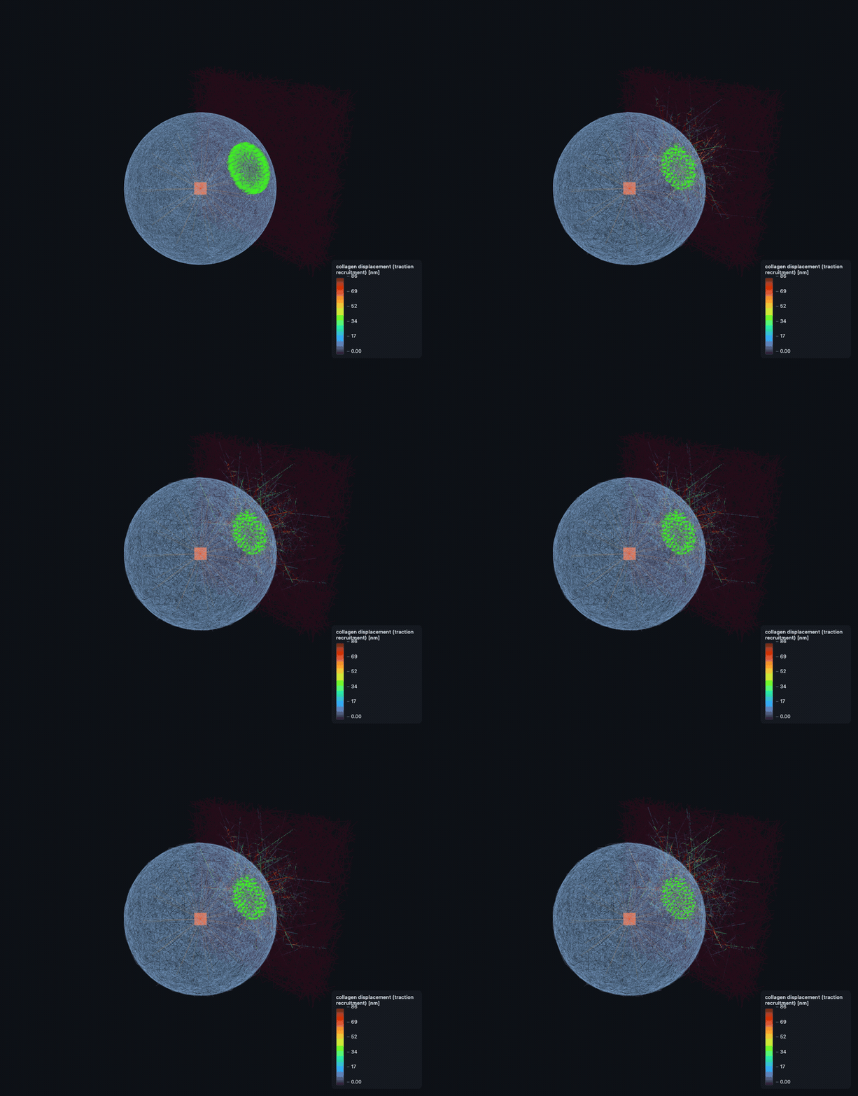
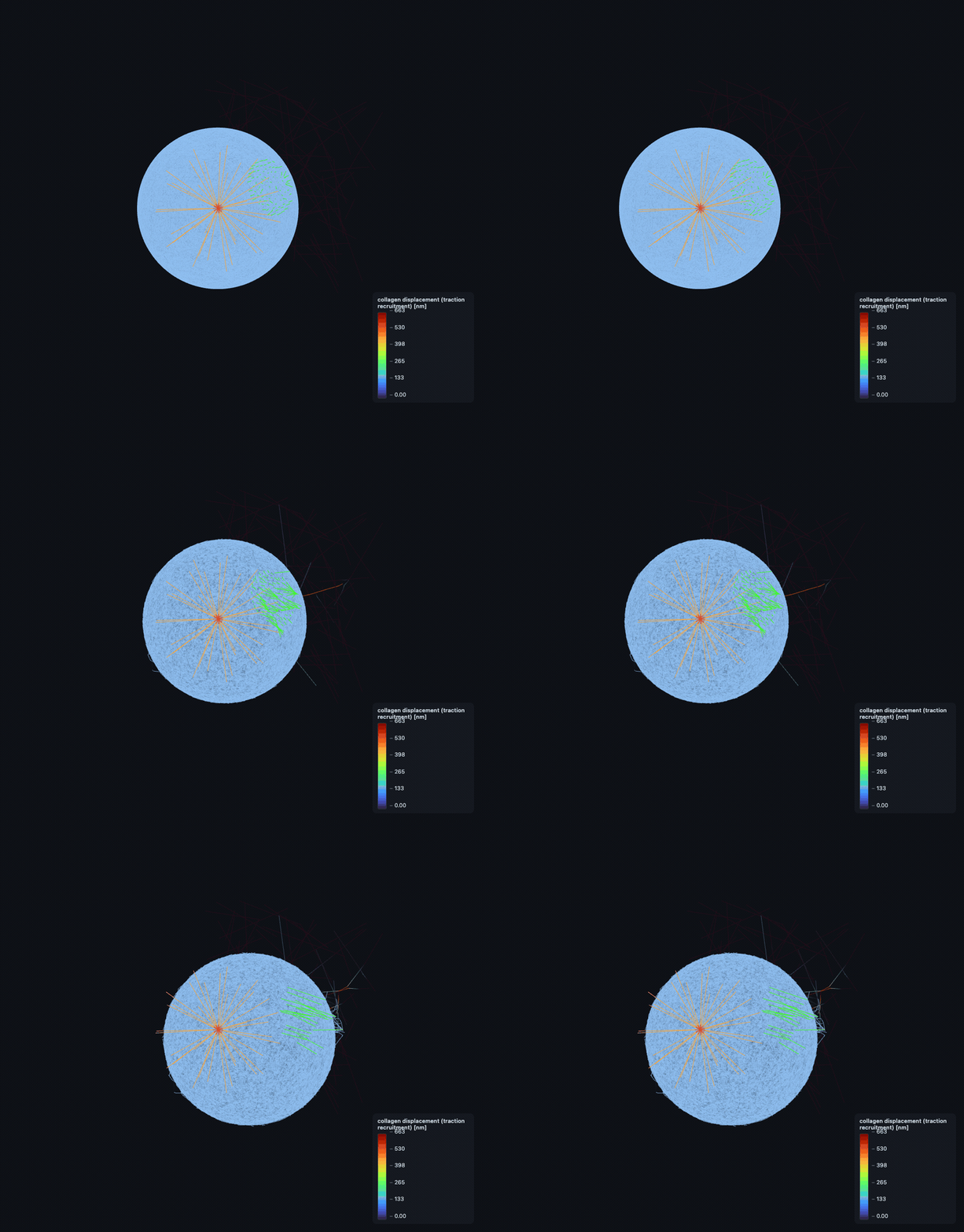
Native FA integrin clutches grip an explicit collagen-I Mikado matrix and contract it — ~+265 nm densification, ~187 nN traction, per-clutch load at the physiological F* — traction-driven and fully two-way.

See the flagship ↓

A 6-material ECM library in real Pascals

Collagen → agarose all validated inside their literature moduli bands; nematic alignment sets E∥/E⊥ from 1× to 63×, and an aligned matrix channels a contractile inclusion's stress far beyond the isotropic case.

Open the ECM viewers ↓

Tissue mechanics from explicit cell–cell junctions

Rakshit cadherin catch-bonds + IPC log-barrier contact give emergent aggregation, faceting, compaction, division and per-cell von-Mises junction stress in confluent spheroids to N=2,000.

See the tissue viewer ↓

Physics implemented — every mechanism, explicit

The heart of the framework: 46 explicit mechanisms across the two engines (29 in FF, 17 in DCM), each a real particle/bond process — paper closed-forms (Bell–Evans, Hill, Pereverzev, WLC/Cytosim bending, Rakshit catch-bonds, IPC) are the runtime mechanism, never a wrapper. Below is a scannable map grouped by subsystem; click any mechanism to expand its modelling detail, its governing equation, a real biological figure of what it is, and its validation/oracle figure. Status: ✓ validated against an oracle · △ honest shortfall (annotated, never hidden) · ⚠ additive / PI-gated · ● core production path · ★ new this cycle.

FF — Filament-FEM engine · the explicit, fine-grained cytoskeleton (Cytosim physics)

Filament mechanicsthe explicit cytoskeleton — bending, stretch, crosslinks, growth

Cytosim WLC bendingsemiflexible filament bending, paper-literal✓ validated
\(\mathbf F_i=\alpha\,(\mathbf m_{i-1}-2\mathbf m_i+\mathbf m_{i+1}),\quad \alpha=\kappa/\ell^3,\ \ \kappa=k_BT\,L_p\)

Discrete Cytosim bending force per interior triple F=α·(m₋−2m₀+m₊) with the {−F,+2F,−F} stencil, α=κ/seg³ and κ=k_BT·L_p; actin L_p=17µm → κ≈0.073 pN·µm². One Warp thread per triple, atomic-add, float64. Anti-lumped: the paper's discrete energy IS the mechanism.

Literature / anchor: Nédélec & Foethke 2007 NJP 9:427 (discrete bending); Gittes 1993 actin L_p=17µm.

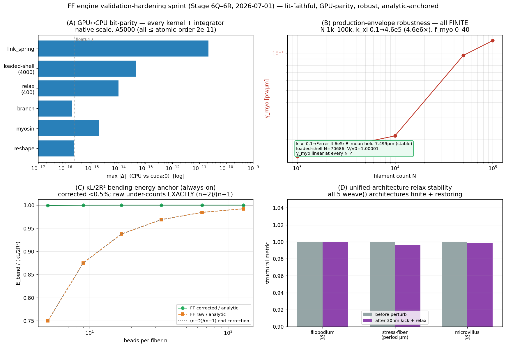
Validation: Cytosim C++ parity oracle on arc bending energy; continuum κL/2R² recovered with the p/(p−1) end-correction (<0.5% error); Euler buckling π²κ/L².

Single-fiber bending energy vs bead count — FF matches the WLC analytic to &lt;0.5%.
validation Single-fiber bending energy vs bead count — FF matches the WLC analytic to <0.5%.
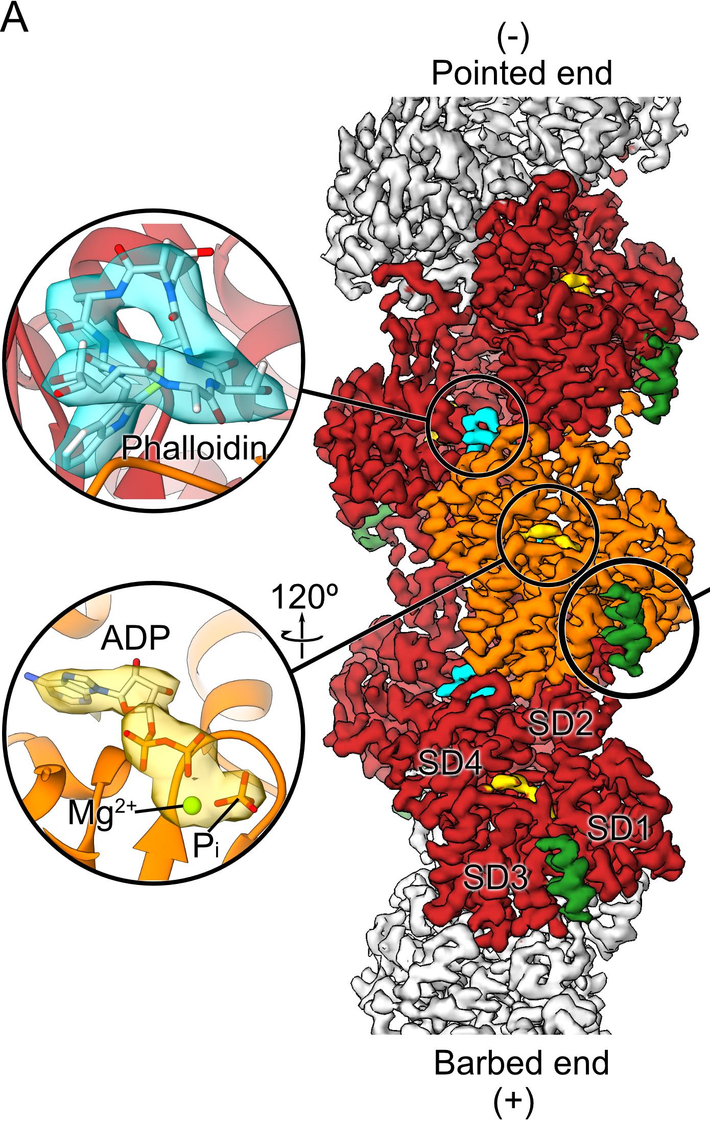
Cryo-EM density map of an F-actin filament — the right-handed double helix of actin subunits (subdomains SD1–SD4) runnin
the biology Cryo-EM density map of an F-actin filament — the right-handed double helix of actin subunits (subdomains SD1–SD4) running from the barbed (+) to the pointed (−) end, the semiflexible polymer the model bends.Belyy et al., PLoS Biol. 2020;18:e3000925 · CC BY 4.0
Fiber inextensibilitya hard constraint, not an axial penalty spring● production path
\(|\mathbf m_{k+1}-\mathbf m_k|=\ell_0,\qquad P=I-J^{\mathsf T}(JJ^{\mathsf T})^{-1}J\)

Segment length fixed by the NF2007 constraint projector P=I−Jᵀ(JJᵀ)⁻¹J plus an exact geometric 'reshape' that restores |mₖ₊₁−mₖ|=seg conserving the centre of mass — explicitly NOT a stretch spring. Reshape exists as both numpy and a one-thread-per-fiber Warp kernel.

Literature / anchor: Nédélec & Foethke 2007 §5.3.

Validation: Pure stretch yields the correct +T tension via the constraint multipliers; reshape restores segment lengths to ~1e-6 µm.

Cryo-EM density map of an F-actin filament — the right-handed double helix of actin subunits (subdomains SD1–SD4) runnin
the biology Cryo-EM density map of an F-actin filament — the right-handed double helix of actin subunits (subdomains SD1–SD4) running from the barbed (+) to the pointed (−) end, the semiflexible polymer the model bends.Belyy et al., PLoS Biol. 2020;18:e3000925 · CC BY 4.0
Crosslinker axial springsα-actinin / filamin cross-fiber Hookean links● production path
\(\mathbf F=k\,(L-r_0)\,\hat{\mathbf u},\qquad k_{\alpha\text{-actinin}}=4.6\times10^5\ \text{pN/µm}\)

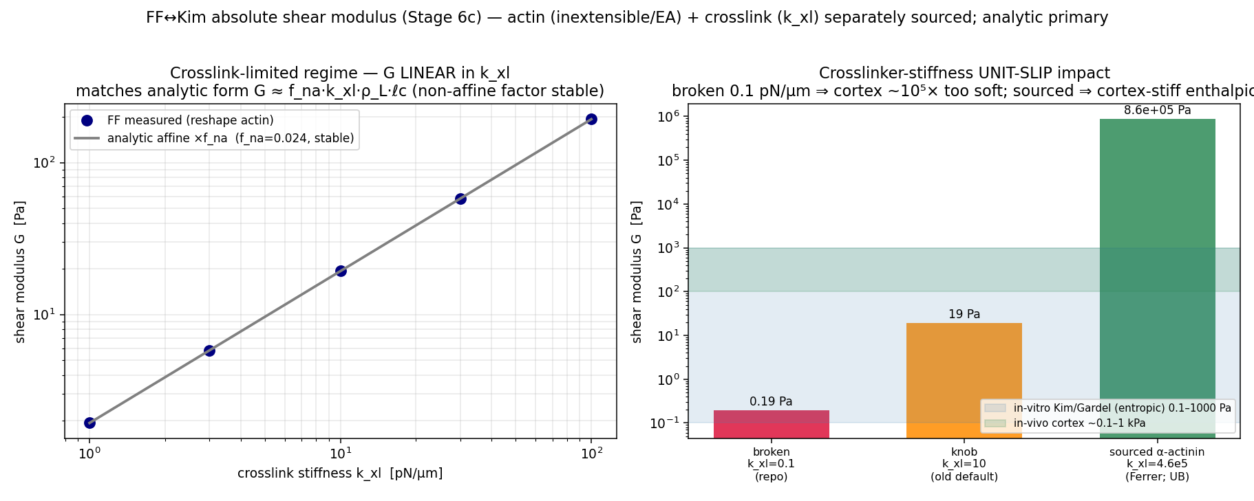
Hookean cross-fiber links k·(L−r₀)·û placed by KDTree near-pairs on different fibers, rest length = formation distance (force-free at build). α-actinin k=4.6e5 pN/µm, filamin k=8.2e5; cortex 30/70 split. Sourced stiffness (not a soft convenience knob).

Literature / anchor: Ferrer 2008 PNAS AFM 455/820 pN/nm; Stricker 2010 30% α-actinin; filamin catch-slip KU-3.19.

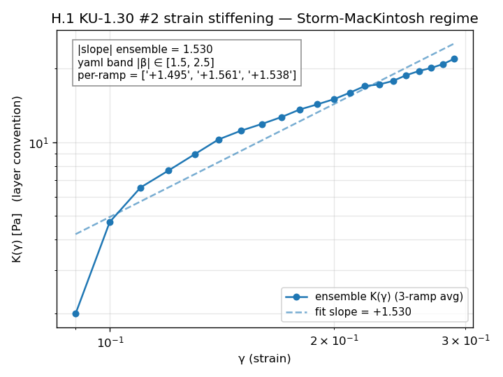
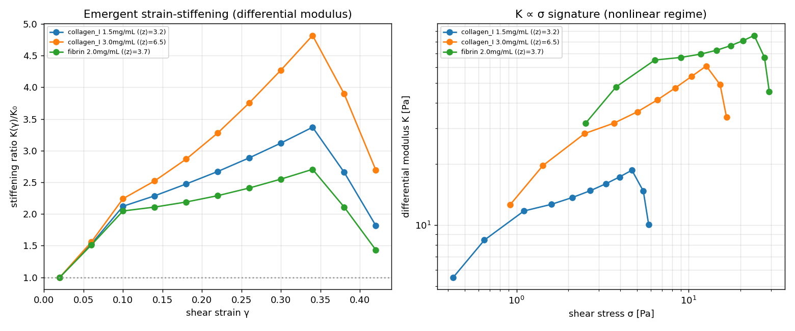
Validation: Bit-parity Warp link-spring kernel vs numpy reference; network strain-stiffening exponent |β|=1.53 sits inside the Storm–MacKintosh [1.5,2.5] band.

Crosslinked-network strain-stiffening exponent β — inside the semiflexible band (lower ~3/2 edge, honest).
validation Crosslinked-network strain-stiffening exponent β — inside the semiflexible band (lower ~3/2 edge, honest).
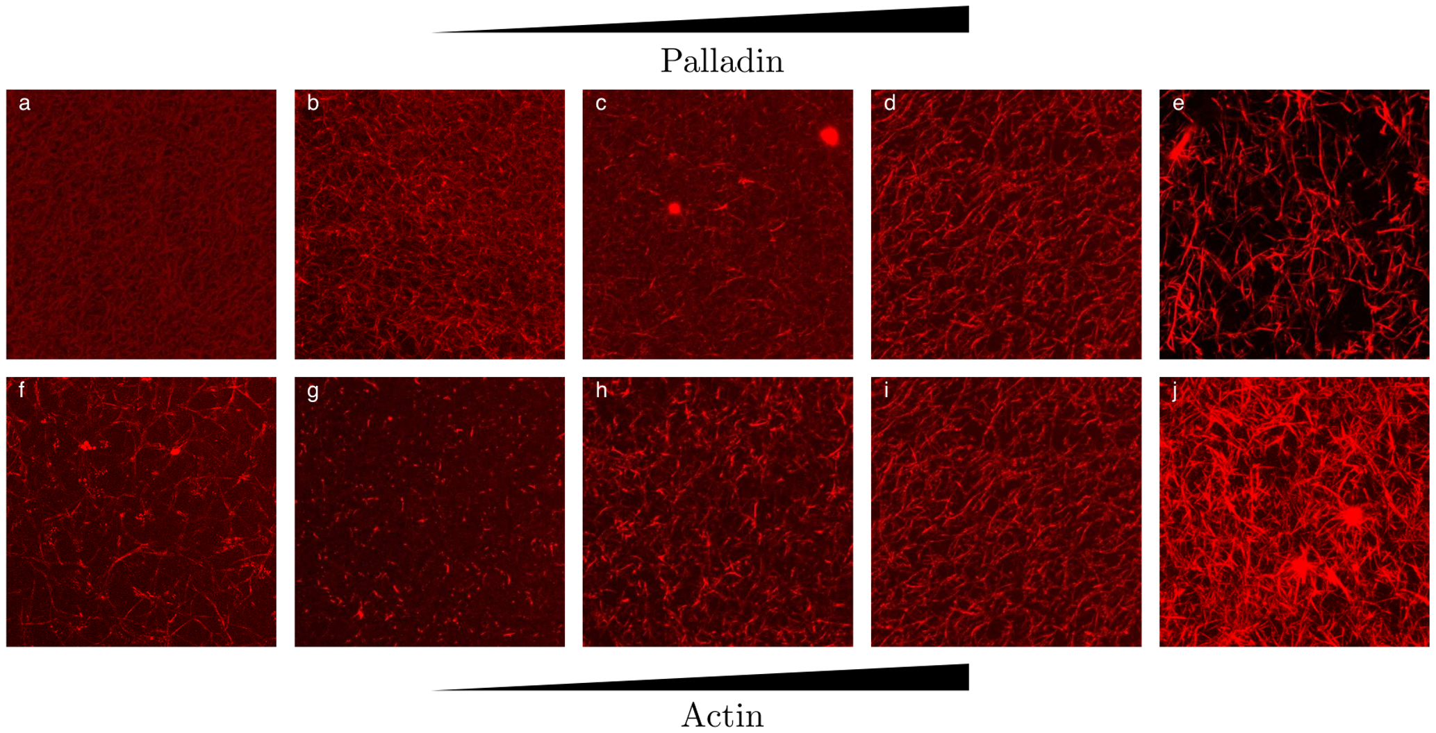
Reconstituted F-actin networks (red, phalloidin) with increasing crosslinker density (a→e): a fine isotropic meshwork tu
the biology Reconstituted F-actin networks (red, phalloidin) with increasing crosslinker density (a→e): a fine isotropic meshwork turns into a coarse bundled network — the crosslinker-driven cortical-actin architecture (crosslinker here is palladin, illustrating the same α-actinin/filamin bundling principle).Grooman et al., PLoS ONE 2012;7:e42773 · CC BY 4.0
Crosslinker viscoelastic turnoverBell-slip Maxwell stress relaxation⚠ additive / PI-gated
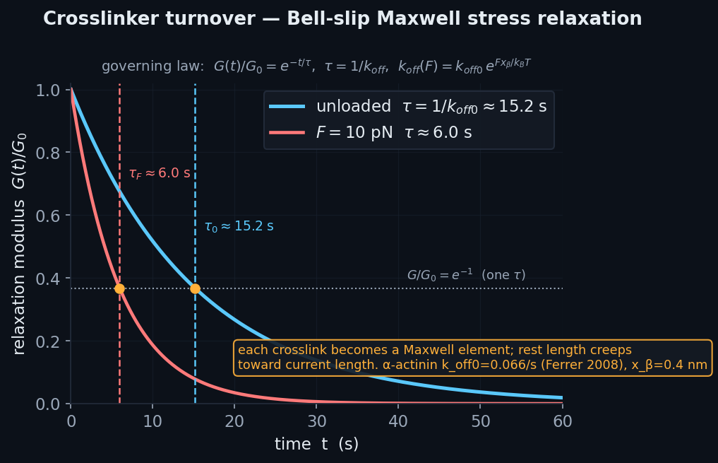
\(k_{\text{off}}(F)=k_{\text{off}}^{0}\,e^{|F|x_\beta/k_BT},\qquad \tau=1/k_{\text{off}}\)

Each turnover event unbinds a fraction 1−exp(−k_off(F)·dt) of the crosslinker ensemble and rebinds force-free at the current geometry, so rest length creeps toward current length — each crosslink becomes a Maxwell element. Off-rate k_off(F)=k_off0·exp(|F|·x_β/kT).

Literature / anchor: NF2007 §10.1 Bell slip; Ferrer 2008 α-actinin k_off0=0.066/s, x_β=0.4nm.

Validation: Mechanism-level (feeds the γ resting baseline); fluidizes the cortex for crawl and binds the stiff crosslinks force-free.

Analytic Bell-slip Maxwell stress relaxation G(t)=G₀·e^(−t/τ), τ=1/k_off (α-actinin τ≈15 s).
the physics Analytic Bell-slip Maxwell stress relaxation G(t)=G₀·e^(−t/τ), τ=1/k_off (α-actinin τ≈15 s).
Reconstituted F-actin networks (red, phalloidin) with increasing crosslinker density (a→e): a fine isotropic meshwork tu
the biology Reconstituted F-actin networks (red, phalloidin) with increasing crosslinker density (a→e): a fine isotropic meshwork turns into a coarse bundled network — the crosslinker-driven cortical-actin architecture (crosslinker here is palladin, illustrating the same α-actinin/filamin bundling principle).Grooman et al., PLoS ONE 2012;7:e42773 · CC BY 4.0
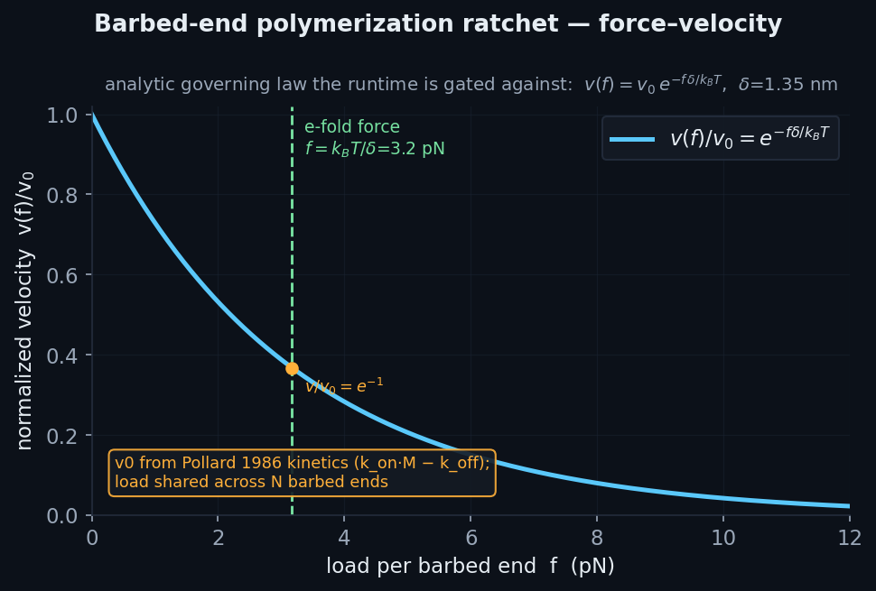
Barbed-end polymerization ratchetload-dependent actin elongation → protrusion✓ validated
\(v(f)=v_0\,e^{-f\,\delta/k_BT},\qquad \delta=1.35\ \text{nm}\)

Load-dependent barbed-end growth v(f)=v₀·exp(−f·δ/k_BT) grown into the tip segment's rest length (reshape advances the tip = protrusion); v₀ from Pollard kinetics, δ=1.35nm half-monomer; membrane load shared across N barbed ends. Filaments grow at their own emergent local load.

Literature / anchor: Mogilner–Oster Brownian ratchet (KU-3.6); Pollard 1986 k_on=11.6/µM/s, k_off=1.4/s.

Validation: Analytic ground truth v(f)=v₀·exp(−f·δ/k_BT) exactly; e-fold force k_BT/δ≈3.2pN reproduced.

Analytic Mogilner–Oster ratchet force–velocity v(f)=v₀·e^(−fδ/kBT) — the closed-form governing law, e-fold load kBT/δ≈3.
the physics Analytic Mogilner–Oster ratchet force–velocity v(f)=v₀·e^(−fδ/kBT) — the closed-form governing law, e-fold load kBT/δ≈3.2 pN marked.
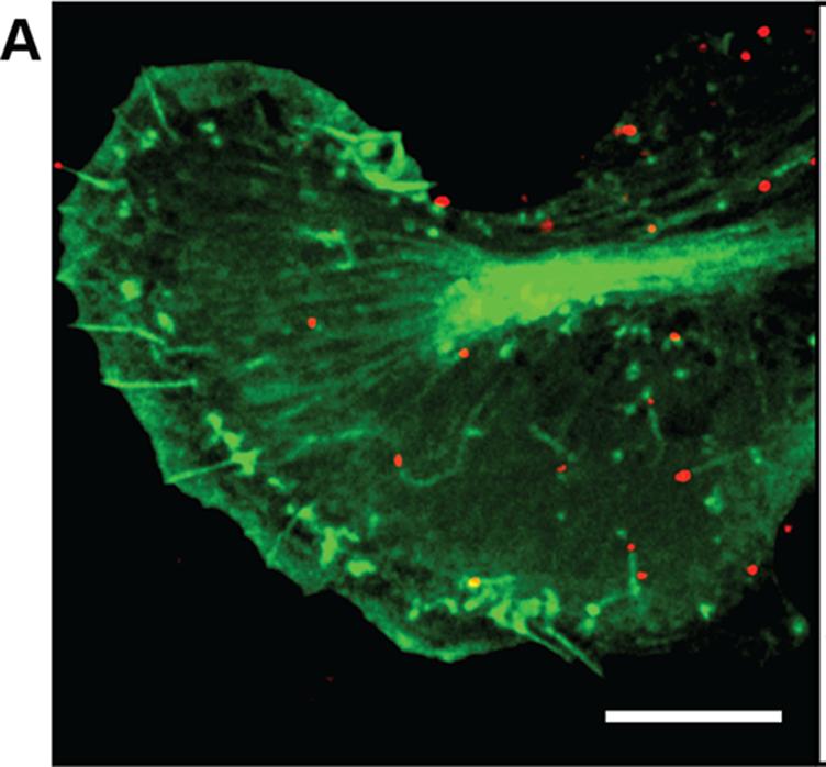
A GFP-actin cell (green) with a virus particle (red) nucleating an actin comet tail — a dense column of polymerizing act
the biology A GFP-actin cell (green) with a virus particle (red) nucleating an actin comet tail — a dense column of polymerizing actin whose barbed-end elongation propels the particle: the polymerization ratchet in action.Mueller et al., PLoS Biol. 2014;12:e1001765 · CC BY 4.0
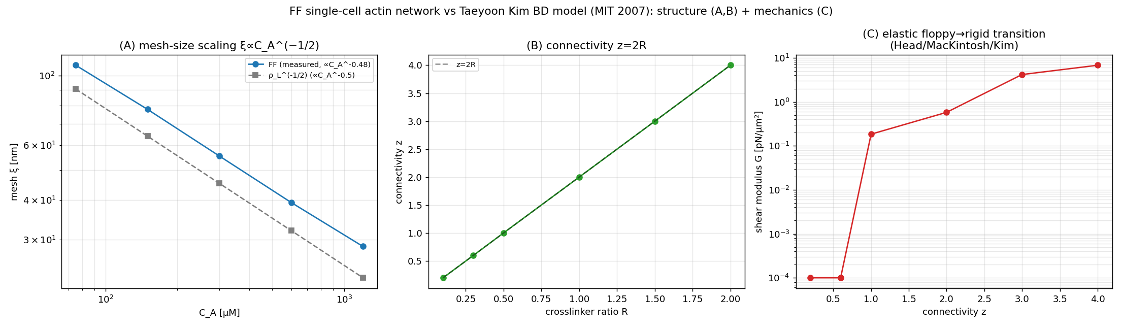
Actin-network structure & rigiditymesh scaling, connectivity, floppy→rigid percolation△ honest shortfall
\(\xi\sim\rho_L^{-1/2},\qquad z=2R\ \ (\text{floppy}\to\text{rigid at isostatic }z)\)

Mesh size ξ~C_A^−0.48 matches the ρ_L^−1/2 law; connectivity z=2R exactly; the elastic floppy→rigid rigidity-percolation transition (G rises from ~0 sub-isostatic through the isostatic point to rigid) is reproduced (Head/MacKintosh/Kim).

Literature / anchor: Taeyoon-Kim Brownian-dynamics actin-network model (Kim 2007).

Validation: Structural/scaling match is exact; the ABSOLUTE modulus inherits the crosslinker-stiffness sourcing tension (shape/trend right, magnitude sourcing-dependent).

Mesh-size scaling, connectivity z=2R, and the floppy→rigid transition — all reproduced.
validation Mesh-size scaling, connectivity z=2R, and the floppy→rigid transition — all reproduced.
Reconstituted F-actin networks (red, phalloidin) with increasing crosslinker density (a→e): a fine isotropic meshwork tu
the biology Reconstituted F-actin networks (red, phalloidin) with increasing crosslinker density (a→e): a fine isotropic meshwork turns into a coarse bundled network — the crosslinker-driven cortical-actin architecture (crosslinker here is palladin, illustrating the same α-actinin/filamin bundling principle).Grooman et al., PLoS ONE 2012;7:e42773 · CC BY 4.0

Motors & contractilityactomyosin prestress — and the honest γ-floor

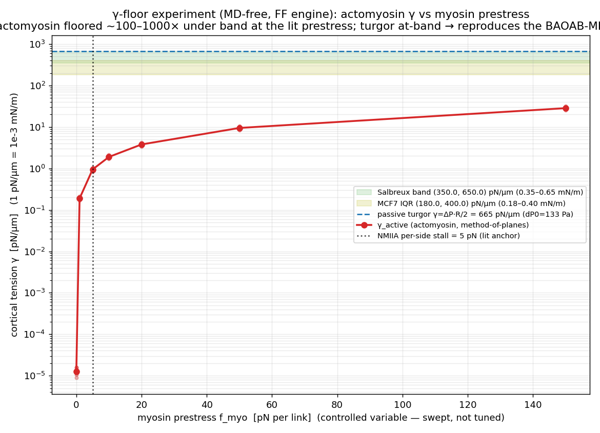
Myosin active prestressthe cortical-tension source (and THE γ-floor)△ honest shortfall
\(\gamma_{\text{myo}}=0.0199\,f_{\text{myo}}\quad(\text{method-of-planes, zero-intercept})\)

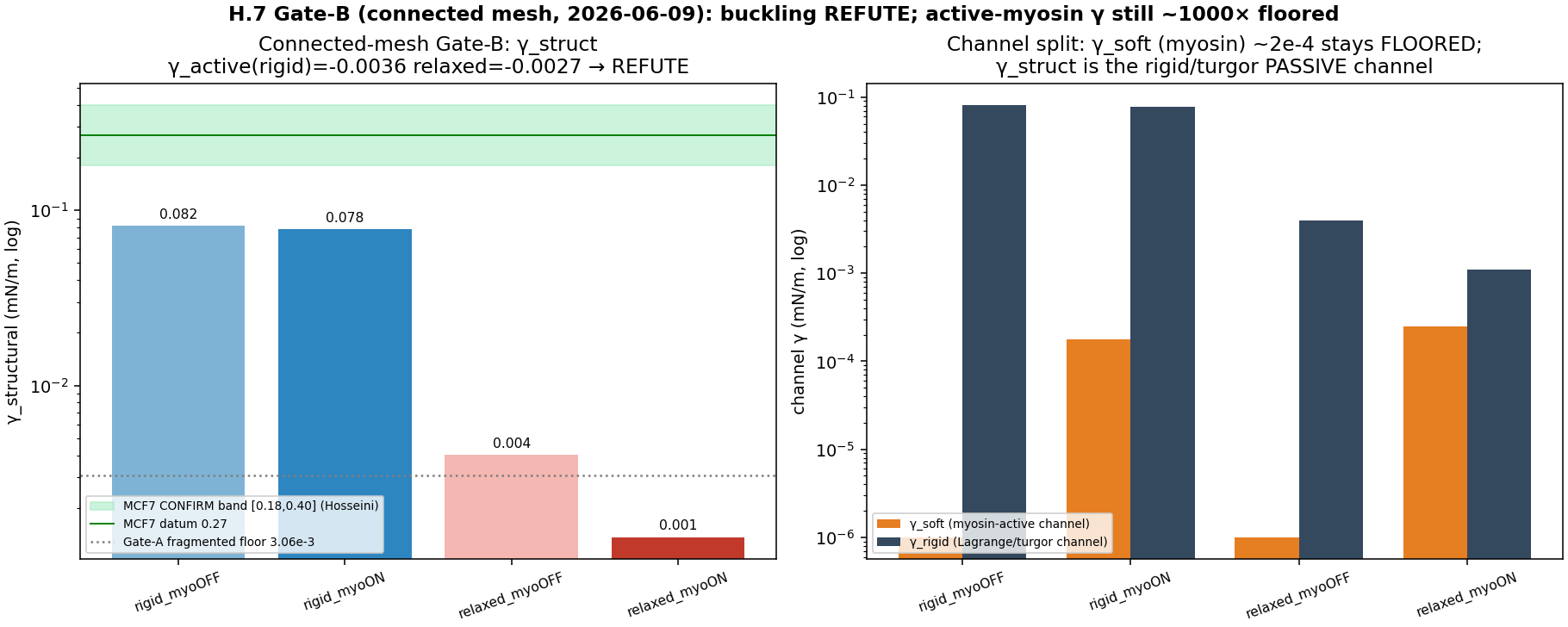
Constant-magnitude contractile dipole f_myo pulling cross-fiber node pairs together, anchored to the NMIIA minifilament stall (0.5 pN/head × ~10 heads/side). This is the actomyosin prestress the method-of-planes γ reads. The honest γ-floor: MD-free active γ_myo runs hundreds-fold under the myosin-active band (~530× at the Nie-sparse production density), robust to buckling/turnover/connectivity/crosslink-stiffness. No MCF7-adherent engaged-NMII density datum exists (open lever).

Literature / anchor: Kovács 2003 F_stall/head=0.5pN; Stam-Hocky/AFINES ~10 heads/side; Nie 2015 areal density 0.625/µm².

Validation: Method-of-planes γ_myo linear in f_myo, zero-intercept (γ=0.0199·f_myo); ensemble quenched realizations.

FF actomyosin γ_active vs myosin prestress — floored ~100–1000× under the MCF7/Salbreux band.
validation FF actomyosin γ_active vs myosin prestress — floored ~100–1000× under the MCF7/Salbreux band.
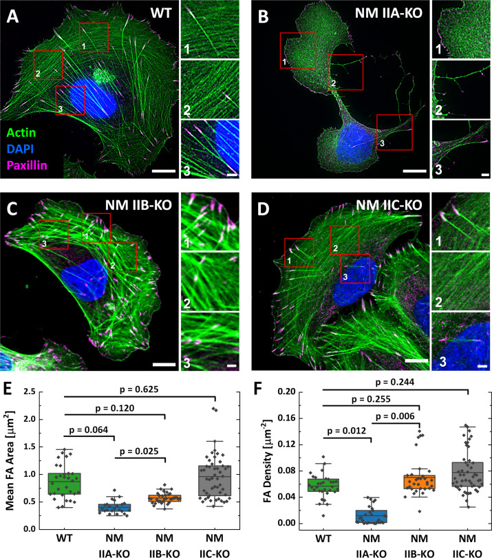
Fluorescence of a cell — F-actin (green), the focal-adhesion marker paxillin (magenta), DNA (blue): the contractile acto
the biology Fluorescence of a cell — F-actin (green), the focal-adhesion marker paxillin (magenta), DNA (blue): the contractile actomyosin stress-fiber array that non-muscle myosin II assembles and tensions, anchored at focal adhesions.Weißenbruch et al., eLife 2021;10:e71888 · CC BY 4.0
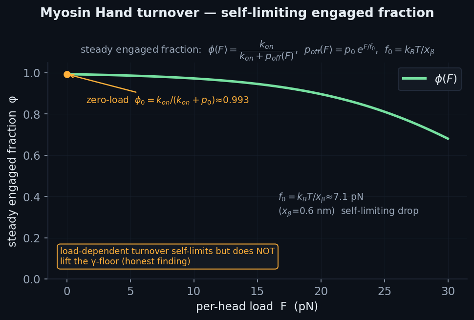
Myosin Hand turnover KMCBell detach / reattach, load self-limiting⚠ additive / PI-gated
\(\phi(F)=\dfrac{k_{\text{on}}}{k_{\text{on}}+p_0\,e^{F/f_0}}\)

Per-link KMC: a bound myosin hand detaches with 1−exp(−τ·p₀·exp(|f|/f₀)) on its own contractile load (Bell slip); a detached hand within capture radius reattaches at k_on. Engaged fraction self-limits under load.

Literature / anchor: NF2007 §10.1 Hand; NMIIA p₀=0.35/s, f₀=kT/x_β (Veigel 2002), k_on=50/s.

Validation: Steady engaged fraction → k_on/(k_on+p_off), τ-independent for small τ. Honest outcome: load-dependent turnover does NOT lift the γ-floor.

Analytic steady engaged fraction φ(F)=k_on/(k_on+p_off(F)) — self-limits but does not lift the γ-floor (honest).
the physics Analytic steady engaged fraction φ(F)=k_on/(k_on+p_off(F)) — self-limits but does not lift the γ-floor (honest).
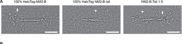
Negative-stain electron micrographs of three bipolar non-muscle myosin II minifilaments — two clusters of myosin heads a
the biology Negative-stain electron micrographs of three bipolar non-muscle myosin II minifilaments — two clusters of myosin heads at opposite ends of a bare central shaft (arrows), the force-generating unit distinct from a whole stress fiber.Melli et al., eLife 2018;7:e32871 · CC0 (public domain)
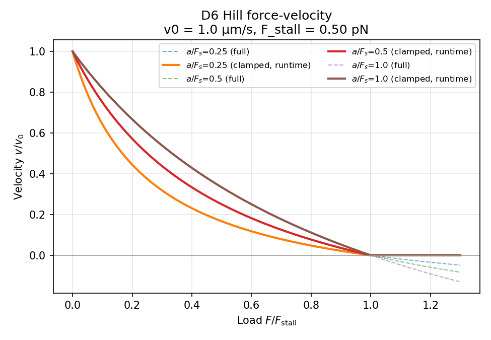
Bipolar minifilamentStam-Hocky explicit multi-head, linear force-velocity⚠ additive / PI-gated
\(v(F)=v_0\,(1-F/F_s),\qquad v_0=0.12\ \text{µm/s}\)

Explicit bipolar minifilament (N_heads=60, per-head stall 2pN, duty 0.1); the contractile force between two actin anchors is DERIVED from the inverted linear force-velocity v(F)=v₀(1−F/Fs), v₀=0.12µm/s. At quasi-static v→0 it gives full stall prestress. PI-ratified: the law is linear, not Hill (Hill 1938 is muscle-only for NMIIA).

Literature / anchor: KB-3.18 composition (N_HEADS=60, F_head=2pN); Kovács 2003 duty=0.1; Freedman 2017 / Tam 2021 linear law.

Validation: Ensemble stall Fs=N_side·F_head in the KB-3.18 50–100pN band; strictly linear FV; force-free at v₀.

Emergent motor-ensemble force-velocity — tracks the Hill/linear oracle to stall, then physically clamps.
validation Emergent motor-ensemble force-velocity — tracks the Hill/linear oracle to stall, then physically clamps.
Negative-stain electron micrographs of three bipolar non-muscle myosin II minifilaments — two clusters of myosin heads a
the biology Negative-stain electron micrographs of three bipolar non-muscle myosin II minifilaments — two clusters of myosin heads at opposite ends of a bare central shaft (arrows), the force-generating unit distinct from a whole stress fiber.Melli et al., eLife 2018;7:e32871 · CC0 (public domain)
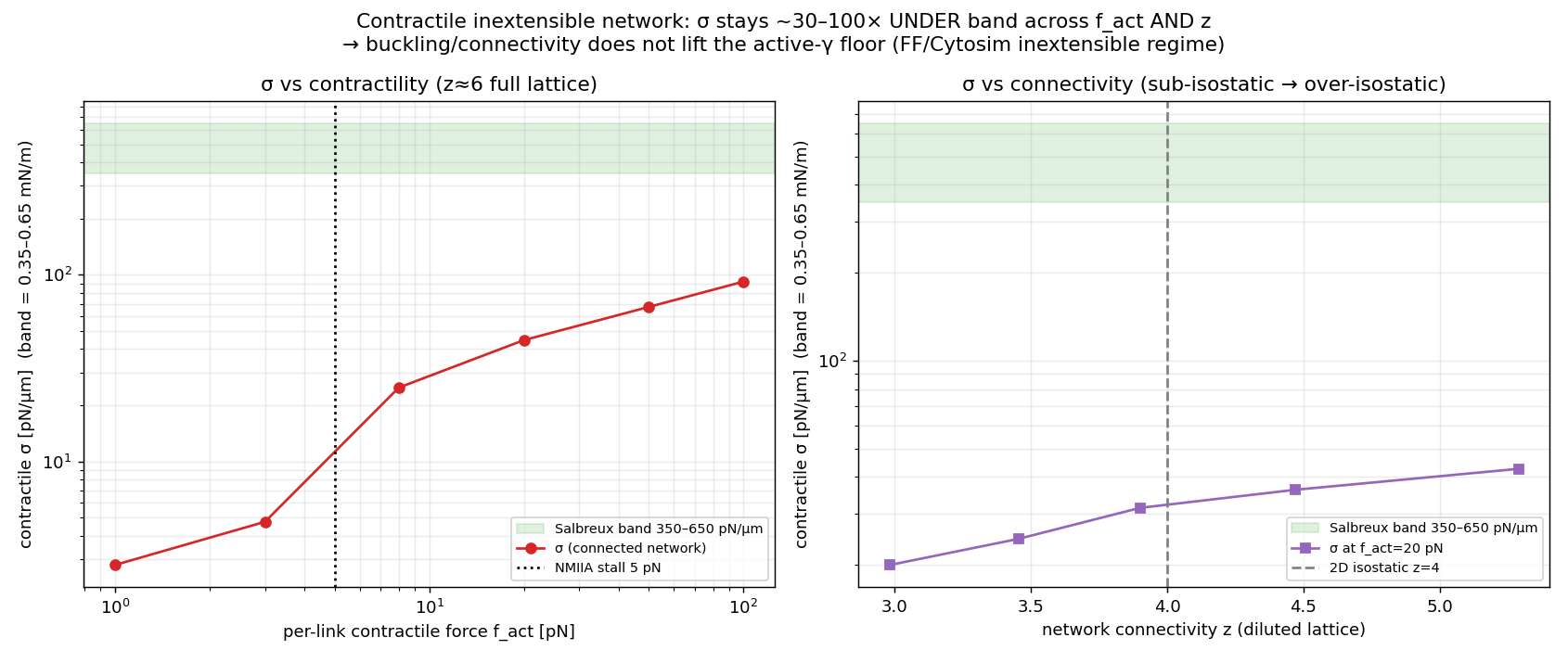
Contractile-network bucklingdoes connectivity lift the γ-floor? (no)△ honest shortfall
\(\sigma(z)\ \text{flat}\ \Rightarrow\ \text{no Ronceray amplification (inextensible)}\)

Connected inextensible contractile network: a 2D triangular lattice of 3-node buckling-capable fibers, edge dilution sweeps the mean coordination z into the sub-isostatic regime, per-fiber contractile motor f_act, boundary pinned; macroscopic σ = boundary reaction / perimeter.

Literature / anchor: Ronceray/Broedersz/Lenz 2016 amplification hypothesis (tested, not assumed).

Validation: Finding: σ stays ~10–90 pN/µm and FLAT in z — buckling/connectivity do NOT lift the γ-floor. Ronceray's ~7× amplification is an EXTENSIBLE-network effect that Cytosim inextensibility structurally precludes.

Network stress vs contractile force and connectivity z — flat in z, confirming the γ-floor is robust.
validation Network stress vs contractile force and connectivity z — flat in z, confirming the γ-floor is robust.
Fluorescence of a cell — F-actin (green), the focal-adhesion marker paxillin (magenta), DNA (blue): the contractile acto
the biology Fluorescence of a cell — F-actin (green), the focal-adhesion marker paxillin (magenta), DNA (blue): the contractile actomyosin stress-fiber array that non-muscle myosin II assembles and tensions, anchored at focal adhesions.Weißenbruch et al., eLife 2021;10:e71888 · CC BY 4.0
Cortical-tension γ estimatormethod-of-planes measurement instrument● production path
\(\gamma=\dfrac{1}{2\pi R}\sum_{\text{cut}} T\,|\hat{\mathbf u}\cdot\hat{\mathbf n}|\)

For Fibonacci diametral planes through the centre, sum T·|û·n̂| of every load-bearing element crossing the plane (actin axial tension + crosslinker links + myosin links) / cut circumference 2πR, averaged. A faithful port of the HOOMD instrument — measured the SAME way as experiment.
Literature / anchor: Archived cortical_tension.py; Chugh 2017 HeLa AFM (350,650); MCF7 Hosseini 180–400 pN/µm.

Validation: The decisive MD-free-vs-BAOAB γ comparison instrument.

Gate-B emergent active cortical tension on the connected ~38k-filament mesh (myoON/OFF, rigid/relaxed).
validation Gate-B emergent active cortical tension on the connected ~38k-filament mesh (myoON/OFF, rigid/relaxed).
Fluorescence of a cell — F-actin (green), the focal-adhesion marker paxillin (magenta), DNA (blue): the contractile acto
the biology Fluorescence of a cell — F-actin (green), the focal-adhesion marker paxillin (magenta), DNA (blue): the contractile actomyosin stress-fiber array that non-muscle myosin II assembles and tensions, anchored at focal adhesions.Weißenbruch et al., eLife 2021;10:e71888 · CC BY 4.0

Adhesion & the molecular clutchintegrin catch-slip, FA maturation, compliant substrate

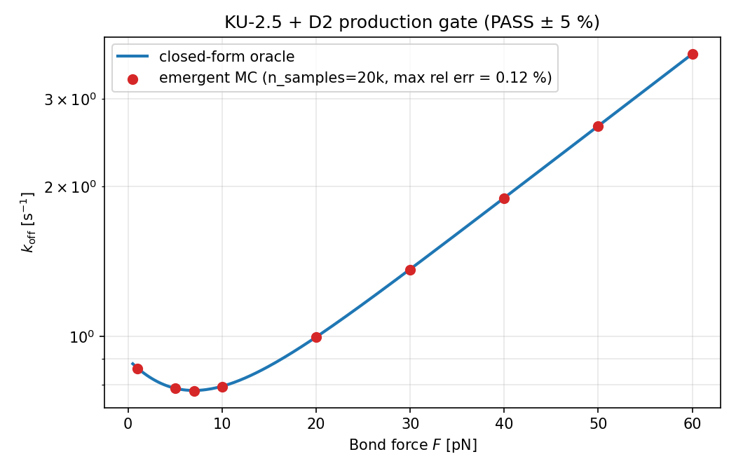
FA integrin clutchmolecular clutch + Pereverzev catch-slip✓ validated
\(k_{\text{off}}(F)=k_{c0}\,e^{-Fx_c/k_BT}+k_{s0}\,e^{+Fx_s/k_BT}\)

Hookean clutch spring from an actin end node to a substrate anchor (traction reaction to the ECM) + Pereverzev two-pathway catch-slip KMC off-rate k_catch0·exp(−Fx_c/kT)+k_slip0·exp(+Fx_s/kT); a bound clutch detaches load-dependently, a detached clutch within capture reattaches at k_on. De-adhesion EMERGENT, never a binary latch.

Literature / anchor: Mitchison-Kirschner 1988 / Chan-Odde 2008 clutch; INTEGRIN_A5B1 Kong 2009 (KB-2.5).

Validation: Kernel validated against the analytic Pereverzev off-rate as-recorded; analytic catch-slip peak F*=kT/(x_c+x_s)·ln[(k_c0x_c)/(k_s0x_s)].

Emergent Monte-Carlo bond off-rate — biphasic catch-slip lifetime, matches the oracle to 0.12%.
validation Emergent Monte-Carlo bond off-rate — biphasic catch-slip lifetime, matches the oracle to 0.12%.
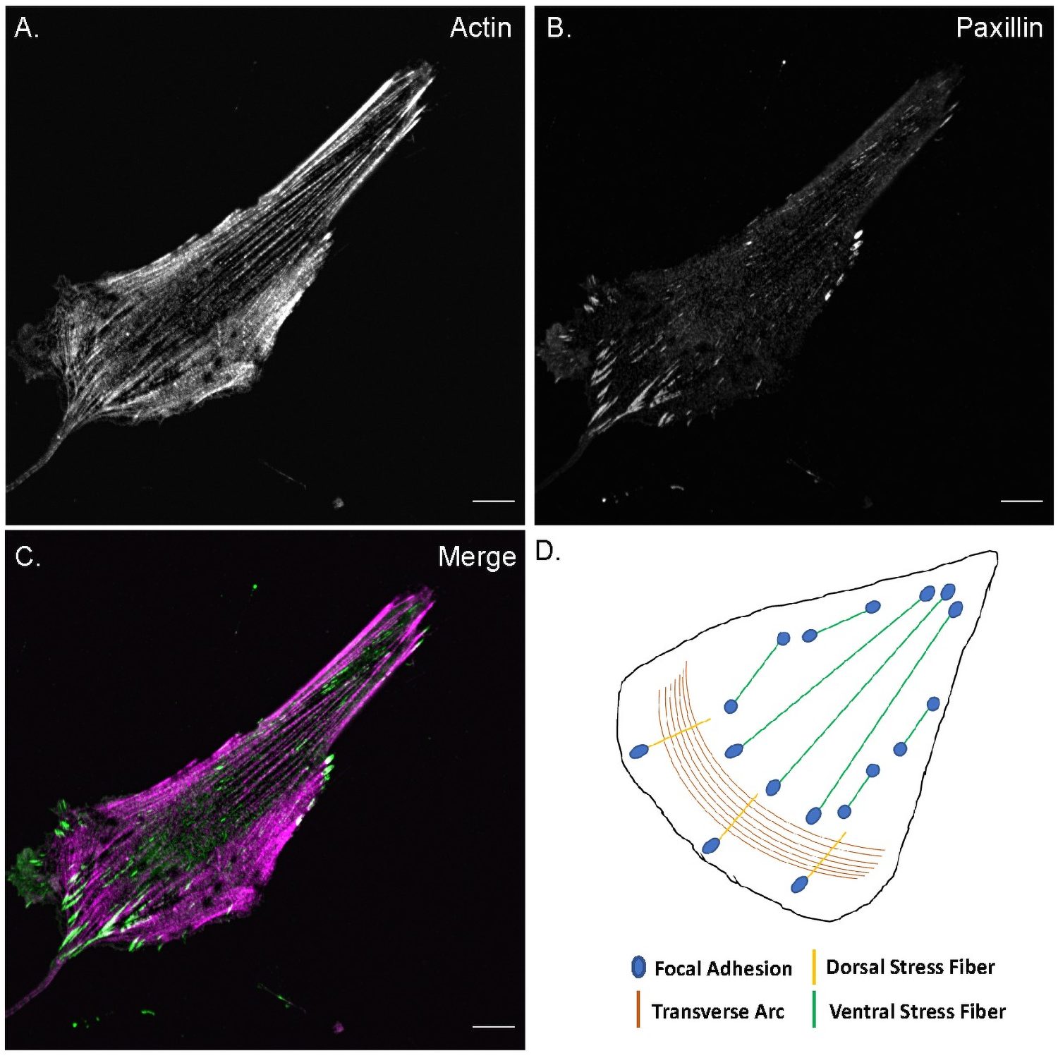
Actin stress fibers (magenta) terminating at focal adhesions marked by paxillin (green puncta), with a schematic of the
the biology Actin stress fibers (magenta) terminating at focal adhesions marked by paxillin (green puncta), with a schematic of the stress-fiber/adhesion organization — the molecular clutches that transmit traction to the ECM.Hauke et al., PLoS ONE 2021;16:e0250749 · CC BY 4.0
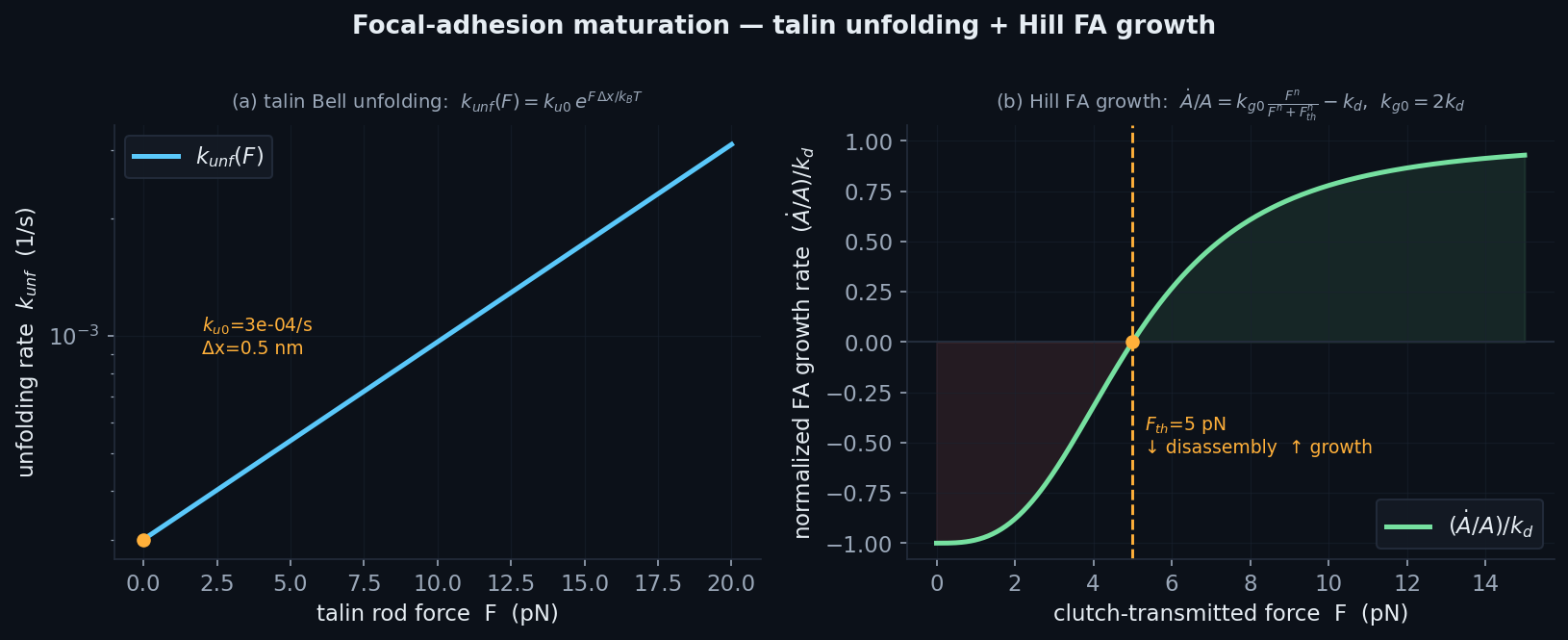
FA maturationtalin unfolding + vinculin + Hill FA-area growth⚠ additive / PI-gated
\(k_{\text{unf}}(F)=k_{u0}\,e^{F\Delta x/k_BT};\quad \dfrac{dA}{dt}=\Big(\dfrac{k_g F^n}{F^n+F_{th}^n}-k_d\Big)A\)

Per-clutch mechanosensor: talin Bell unfolding exposes vinculin sites (force-suppressed refolding); vinculin recruitment reinforces stiffness k_int_eff=k_int·(1+α·N_vin); Hill FA-area growth dA/dt that disassembles below F_th. Force-strengthening adhesion, mechanistically.

Literature / anchor: KB-2.6 talin R3 (F_th=5pN); KB-2.7 vinculin; KB-2.17 Hill FA growth (n=3).

Validation: Talin Bell parity at 5pN; Hill fixed point dA/dt=0 exactly at F=F_th (self-consistent k_g0=2k_d).

Analytic talin Bell unfolding + Hill FA-growth fixed point at F_th=5 pN (below → disassembly, above → growth).
the physics Analytic talin Bell unfolding + Hill FA-growth fixed point at F_th=5 pN (below → disassembly, above → growth).
Actin stress fibers (magenta) terminating at focal adhesions marked by paxillin (green puncta), with a schematic of the
the biology Actin stress fibers (magenta) terminating at focal adhesions marked by paxillin (green puncta), with a schematic of the stress-fiber/adhesion organization — the molecular clutches that transmit traction to the ECM.Hauke et al., PLoS ONE 2021;16:e0250749 · CC BY 4.0
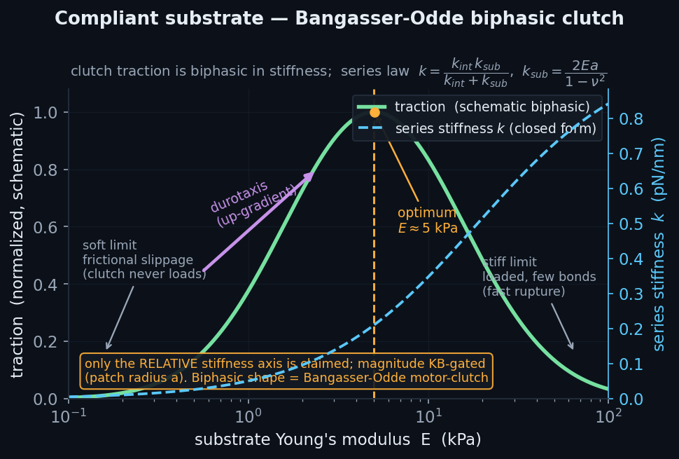
Compliant elastic substrateBangasser-Odde biphasic motility clutch + durotaxis⚠ additive / PI-gated
\(k=\dfrac{k_{\text{int}}\,k_{\text{sub}}}{k_{\text{int}}+k_{\text{sub}}},\qquad k_{\text{sub}}=\dfrac{2Ea}{1-\nu^2}\)

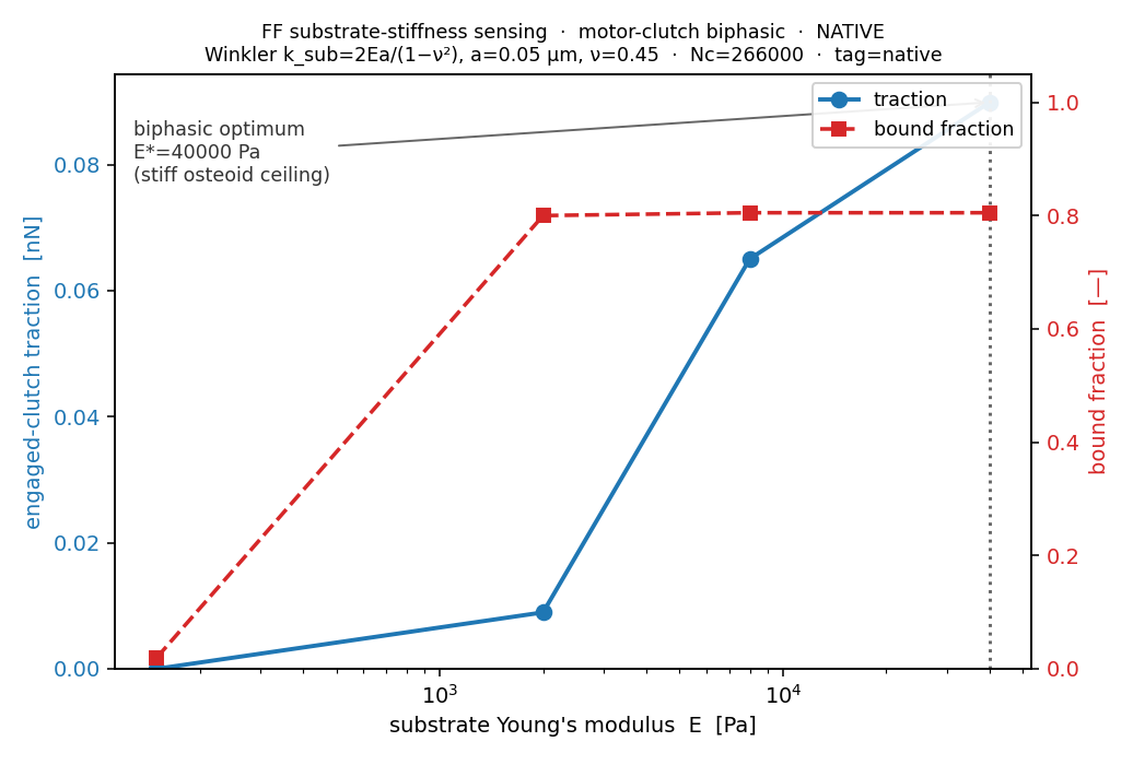
Each FA anchor is a movable point tied to the dish by a Winkler spring k_sub=2Ea/(1−ν²) (Sneddon flat-punch); the actin feels the series stiffness k_int·k_sub/(k_int+k_sub), so traction becomes E-dependent (biphasic) and a stiffness gradient drives durotaxis — emergent from the clutches.

Literature / anchor: KB-1.5 E=5kPa/ν=0.45 PAA; Bangasser-Odde compliant substrate.

Validation: Rigid limit E→∞ recovers the fixed-anchor path byte-for-byte; the RELATIVE stiffness axis (biphasic shape, durotaxis sign) is claimed, magnitude KB-gated.

Analytic Bangasser–Odde biphasic clutch: traction vs substrate stiffness with an optimum; durotaxis up-gradient (relativ
the physics Analytic Bangasser–Odde biphasic clutch: traction vs substrate stiffness with an optimum; durotaxis up-gradient (relative axis only).
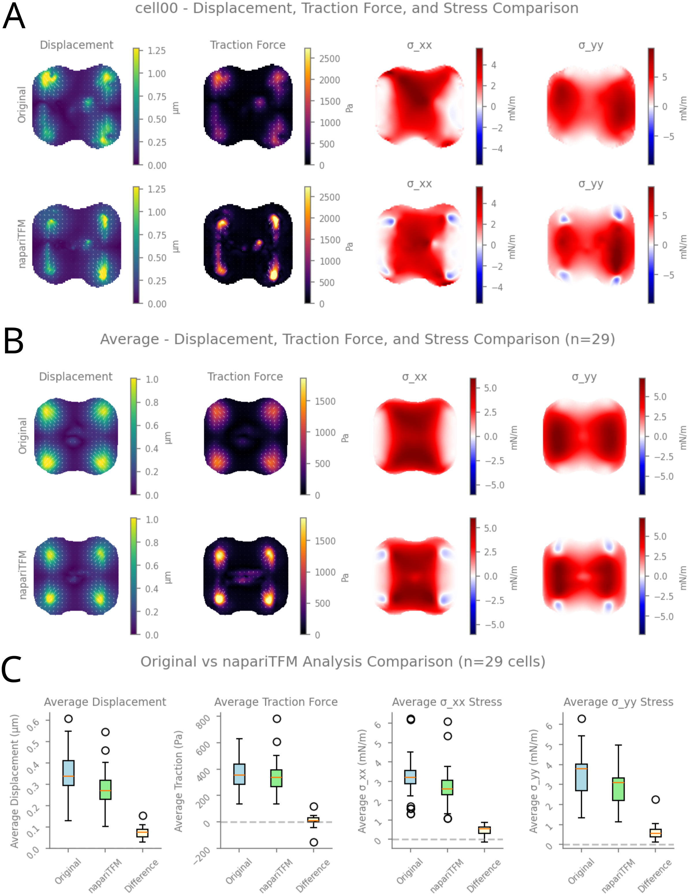
Traction-force microscopy of a single adherent cell: the substrate bead-displacement field and reconstructed traction-st
the biology Traction-force microscopy of a single adherent cell: the substrate bead-displacement field and reconstructed traction-stress map (concentrated at the adhesive corners) — the forces a cell transmits to its elastic substrate.Ruppel et al., PLoS Comput. Biol. 2026;22:e1014045 · CC BY 4.0

Membrane · nucleus · microtubules · cytoplasmthe other compartments, each an explicit body

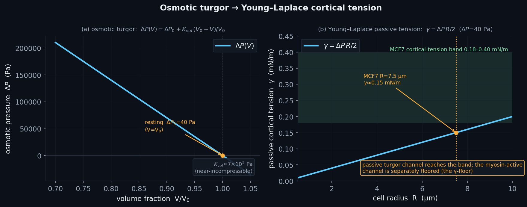
Osmotic turgor / cytoplasmGuo van't Hoff pressure, K_vol derived● production path
\(\Delta P(V)=\Delta P_0+K_{\text{vol}}\dfrac{V_0-V}{V_0},\qquad \gamma_{\text{pass}}=\tfrac12\Delta P\,R\)

State-dependent van't Hoff osmotic pressure ΔP(V) applied as a radial outward Young-Laplace force; the bulk modulus K_vol≈7e5 Pa is DERIVED from c_osm=200mM and V_min=0.30V₀ — no magic K_vol. The passive γ=ΔP·R/2 is the pressure's partner, kept diagnostic-only (never double-booked into actomyosin γ).

Literature / anchor: Guo 2017 PNAS excluded-volume; ΔP₀=40 Pa Fischer-Friedrich 2014; Venkova 2022 V_min=0.30.

Validation: ΔP(V₀)=ΔP₀; K_vol derived; CFL includes the k_turgor breathing mode.

Analytic osmotic-turgor ΔP(V) and its Young–Laplace partner γ=ΔP·R/2 — passive channel reaches the MCF7 band; myosin-act
the physics Analytic osmotic-turgor ΔP(V) and its Young–Laplace partner γ=ΔP·R/2 — passive channel reaches the MCF7 band; myosin-active is separately floored.
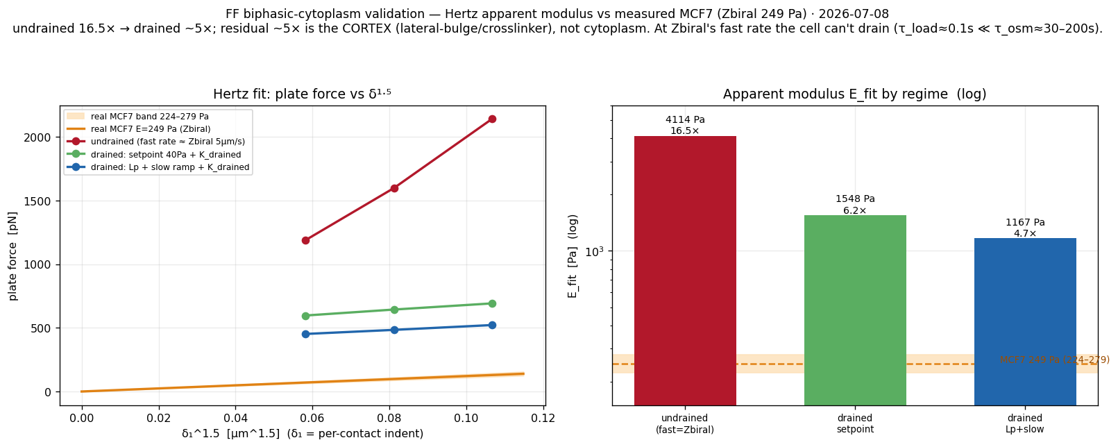
Biphasic poroelastic cytoplasmdrainage + drained solid (Terzaghi)⚠ additive / PI-gated
\(\Delta P=\Delta P_{\text{osm}}+K_{\text{drained}}\max\!\Big(0,\dfrac{V_0-V}{V_0}\Big)\)

(A) Kedem-Katchalsky drainage: the osmotic water reference relaxes toward the geometric volume with a membrane clock τ_osm (analytic, unconditionally stable); (B) Terzaghi effective stress ΔP_solid on the fixed solid rest volume. Additive/default-off — recovers pure turgor bit-identically.

Literature / anchor: Lp=1.6e-8 µm/(s·Pa) COS-7; K_drained≈300 Pa Moeendarbary 2013.

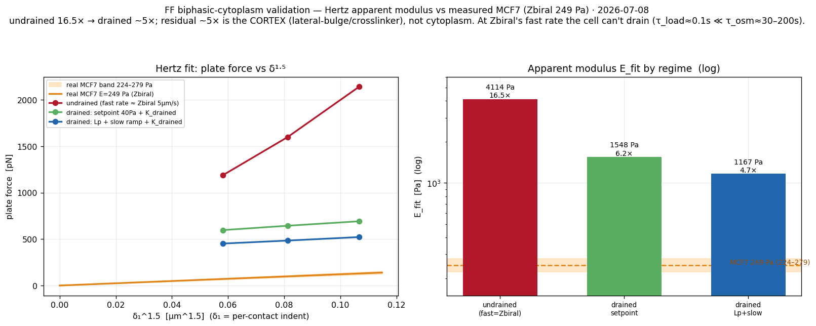
Validation: Poroelastic drainage recovers most of the AFM stiffness gap (τ_load≪τ_osm); a ~5× residual remains, attributed (open) to the cortex not the cytoplasm.

Biphasic-cytoplasm AFM — undrained 16.5× drains to ~4.7× vs measured MCF7 (Zbiral 249 Pa); honest residual.
validation Biphasic-cytoplasm AFM — undrained 16.5× drains to ~4.7× vs measured MCF7 (Zbiral 249 Pa); honest residual.
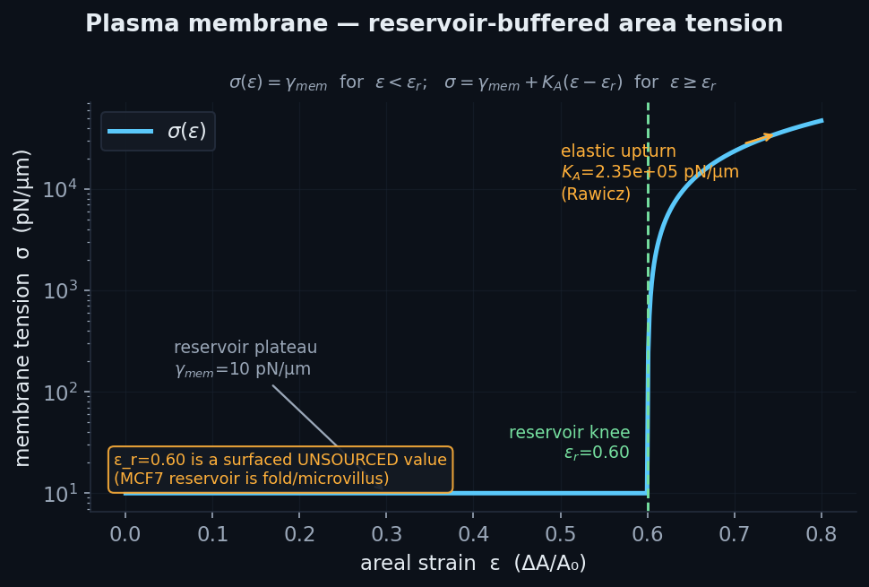
Plasma membraneindependent lipid sheet, reservoir-buffered tension⚠ additive / PI-gated
\(\Delta P=\dfrac{2\gamma}{R},\qquad \gamma=\gamma_{\text{mem}}+K_A\dfrac{S-A_0}{A_0}\)

A separate closed triangulated icosphere with in-plane area-tension (constant γ_mem until the reservoir strain, then a K_A elastic upturn to lysis); breakable ERM tethers to the cortex (rupture → bleb emergent); a one-sided containment penalty stops filament tips leaking out.

Literature / anchor: KU-3.B1: T_m=10 pN/µm (KB-3.B1.1); K_A=2.35e5 Rawicz 2000; ERM tether KB-3.B1.4.

Validation: Warp kernels; delegates params to compartments.resolve_membrane. (RESERVOIR_STRAIN=0.60 surfaced as unsourced.)

Analytic reservoir-buffered membrane tension: constant γ_mem to the strain knee ε_r, then K_A elastic upturn (ε_r=0.60 f
the physics Analytic reservoir-buffered membrane tension: constant γ_mem to the strain knee ε_r, then K_A elastic upturn (ε_r=0.60 flagged unsourced).
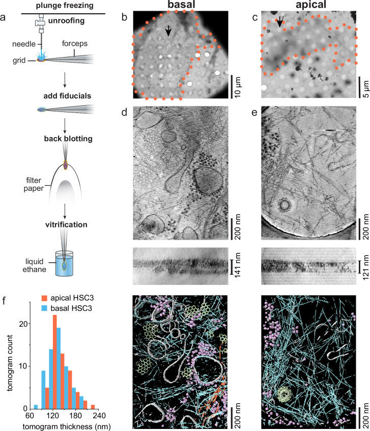
Cryo-electron tomography of an unroofed mammalian plasma membrane and the cortical actin meshwork beneath it (cyan = cor
the biology Cryo-electron tomography of an unroofed mammalian plasma membrane and the cortical actin meshwork beneath it (cyan = cortical filaments) — the membrane–cortex the model treats as an independent lipid sheet on the cortex.Sun et al., Nat. Commun. 2025;16:855 · CC BY 4.0
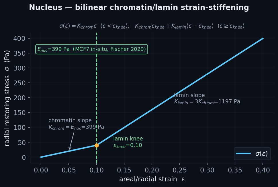
Nucleus (radial shell + envelope)bilinear chromatin/lamin core, real closed surface● production path
\(\sigma(\varepsilon)=\begin{cases}K_{\text{chrom}}\varepsilon & \varepsilon<\varepsilon_k\\ K_{\text{chrom}}\varepsilon_k+K_{\text{lamin}}(\varepsilon-\varepsilon_k) & \varepsilon\ge\varepsilon_k\end{cases}\)

Bilinear strain-stiffening nucleus: soft chromatin slope inside the lamin-engagement knee, stiff lamin slope past it. Two forms — a radial bead-cloud shell and a real closed triangulated nuclear envelope with per-face EMERGENT rupture when areal strain exceeds threshold. Coupled to the cortex by incompressible-volume displacement and direct plate compression.

Literature / anchor: KB-3.B2: E_nuc=399 Pa MCF7 in-situ (Fischer 2020, PI-ratified); RATIO_LAMIN=3.

Validation: law-0 radial-shell parity ~1e-13 vs numpy reference; envelope tangent-modulus knee jump; rupture emerges above / absent below threshold.

Analytic bilinear chromatin→lamin strain-stiffening (E_nuc=399 Pa MCF7, knee 0.10, ×3 lamin slope).
the physics Analytic bilinear chromatin→lamin strain-stiffening (E_nuc=399 Pa MCF7, knee 0.10, ×3 lamin slope).
3D-SIM super-resolution image of the nuclear lamina — the lamin meshwork resolves as a dense fibrous network enclosing g
the biology 3D-SIM super-resolution image of the nuclear lamina — the lamin meshwork resolves as a dense fibrous network enclosing gaps, the stiff strain-stiffening shell around the nucleus.Kittisopikul et al., Cells 2019;8:361 · CC BY 4.0
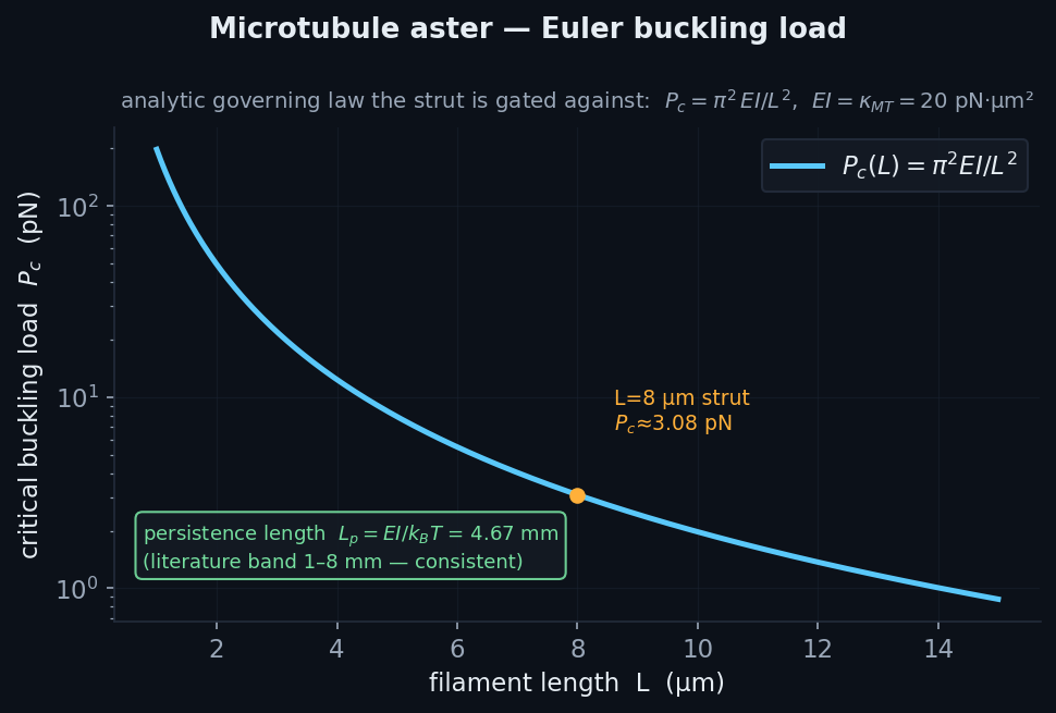
Microtubule aster + MTOC2nd cytoskeletal network, tensegrity strut✓ validated
\(P_c=\dfrac{\pi^2 EI}{L^2},\qquad EI=\kappa_{\text{MT}}=20\ \text{pN·µm}^2\)

Stiff hollow tubes = FF fibers with κ=20 pN·µm² (~300× actin), radiating on Fibonacci directions from one MTOC node held by stiff hub crosslinks; same Cytosim bending + reshape. Compression load-bearing EMERGES from the bending term resisting Euler buckling.

Literature / anchor: Nédélec & Foethke 2007 EI=20; Gittes 1993 cross-check.

Validation: VERIFIED: discrete buckling load converges to Euler π²EI/L² (5.0→1.3% as segments refine); L_p=EI/k_BT=4.67mm in the [1,8]mm band.

Analytic Euler critical load P_c=π²EI/L² (EI=20 pN·µm²) — L_p=EI/kBT=4.67 mm sits in the 1–8 mm band.
the physics Analytic Euler critical load P_c=π²EI/L² (EI=20 pN·µm²) — L_p=EI/kBT=4.67 mm sits in the 1–8 mm band.
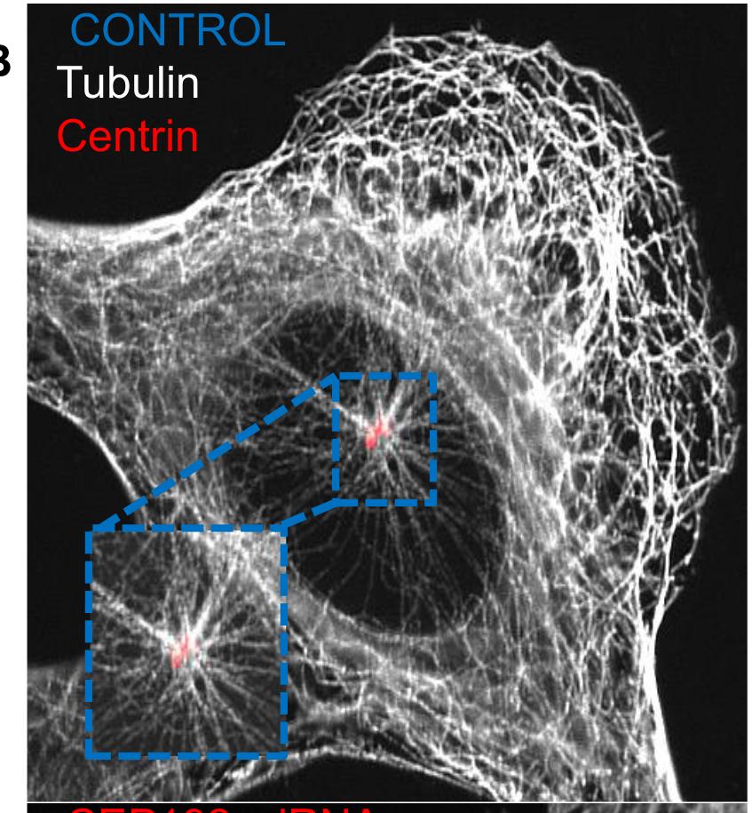
Immunofluorescence of an interphase cell: microtubules (white) radiate from the centrosomal MTOC (centrin, red) as a rad
the biology Immunofluorescence of an interphase cell: microtubules (white) radiate from the centrosomal MTOC (centrin, red) as a radial aster — the stiff second cytoskeletal network that bears compression as a tensegrity strut.O'Rourke et al., PLoS ONE 2014;9:e101001 · CC BY 4.0
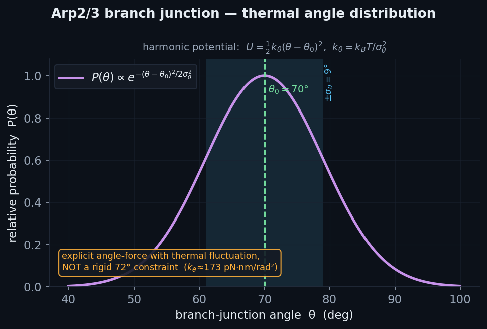
Arp2/3 branch junctionangle-harmonic with thermal fluctuation✓ validated
\(U=\tfrac12 k_\theta(\theta-\theta_0)^2,\qquad \theta_0=70^\circ,\ \sigma_\theta\approx9^\circ\)

Harmonic angle force U=½k_angle·(θ−θ₀)² at each mother-daughter branch triple; θ₀=70°, k_angle=kT/Var(θ) with σ_θ≈9° (equipartition). An explicit angle-force stencil with thermal spread, NOT a rigid 72° constraint.

Literature / anchor: Cryo-ET 68±9° branch angle; Arp2/3 geometry Magic-Number-Blocked.

Validation: Bit-parity Warp branch-angle kernel vs pure-numpy reference.

Analytic equipartition Gaussian branch-angle distribution (θ₀=70°, σ≈9°) — a thermal oracle, not a rigid 72° constraint.
the physics Analytic equipartition Gaussian branch-angle distribution (θ₀=70°, σ≈9°) — a thermal oracle, not a rigid 72° constraint.
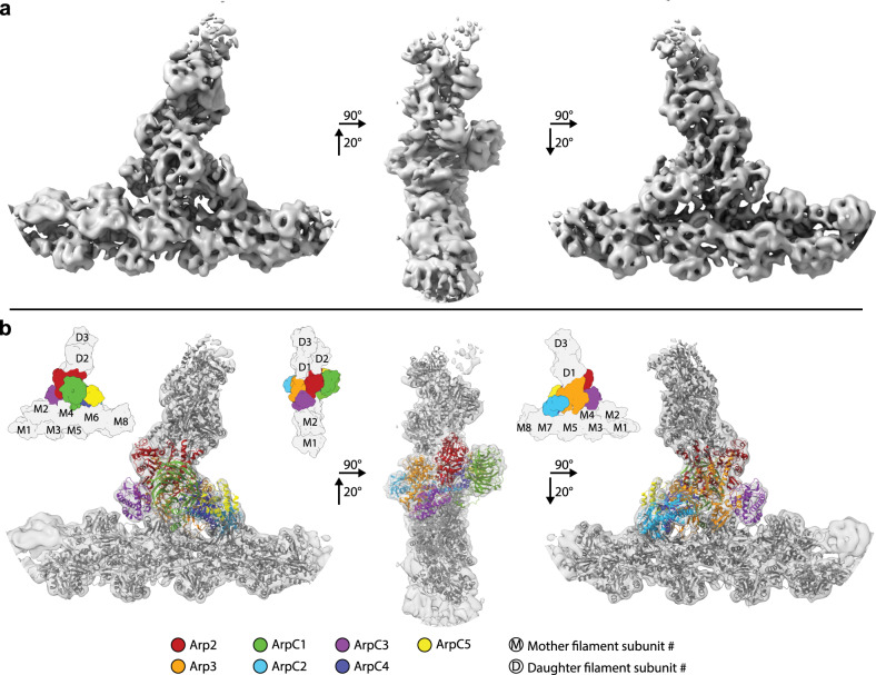
In-cell cryo-electron tomography of the actin–Arp2/3 branch junction: the Arp2/3 complex binds a mother filament and nuc
the biology In-cell cryo-electron tomography of the actin–Arp2/3 branch junction: the Arp2/3 complex binds a mother filament and nucleates a daughter at the characteristic ~70° angle — the building block of the dendritic branched-actin network.Fäßler et al., Nat. Commun. 2020;11:6437 · CC BY 4.0
Excluded-volume soft contactone-sided steric penalty⚠ additive / PI-gated
\(\mathbf F=k_c\,(r_c-L)\,\hat{\mathbf u}\quad[L

One-sided repulsive penalty k·(r_contact−L)·û only when L<r_contact (no attraction) between specified node pairs — used for microtubule-tip↔cortex and filopodium-tip↔ECM-fiber, so the load is supplied EMERGENTLY by the network, not imposed.

Literature / anchor: Generic steric contact (Ericson-style penalty).

Validation: Mechanism-level penalty force law.

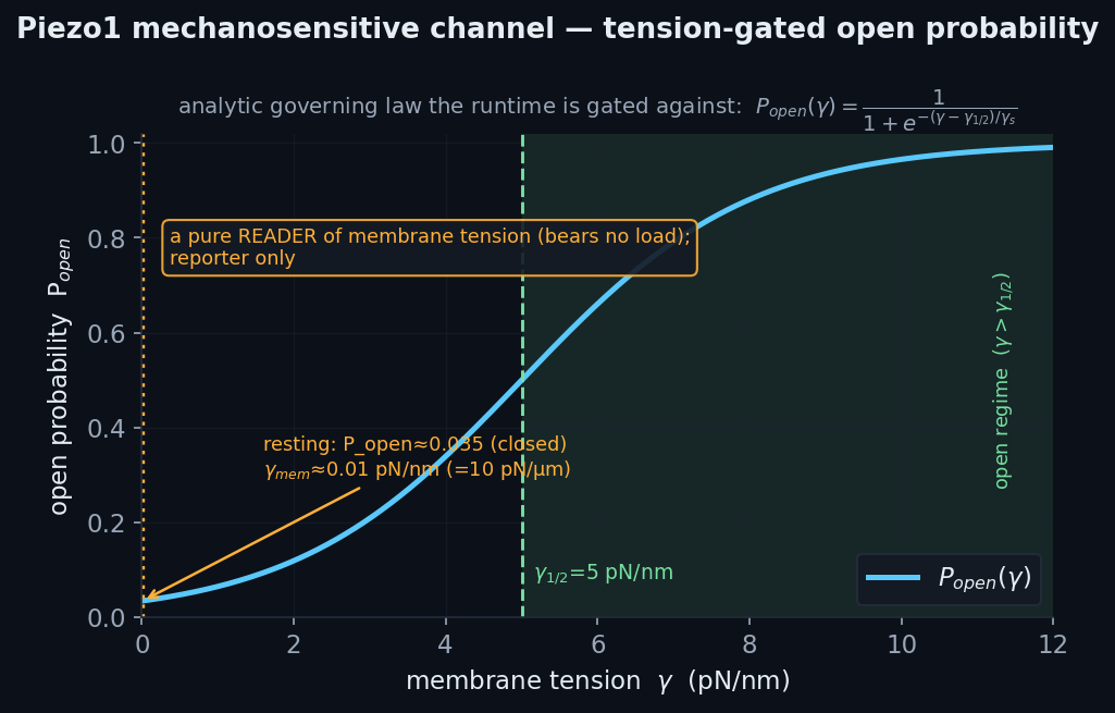
Piezo1 mechanosensitive channeltension-gated open-probability reporter⚠ additive / PI-gated
\(P_{\text{open}}(\gamma)=\dfrac{1}{1+e^{-(\gamma-\gamma_{1/2})/\gamma_s}}\)

Tension-gated open probability P_open(γ)=1/(1+exp(−(γ−γ½)/γ_s)); a pure READER of the membrane tension the model already computes (bears no mechanical load), area-weighted to a whole-cell open fraction. Ca²⁺ feedback is a default-off stub.

Literature / anchor: KB-3.10 Cox 2016 / Lewis-Grandl patch-tension gating.

Validation: Guard gate: at resting γ_mem P_open≈0.035 (closed) — catches the pN/µm vs pN/nm unit trap.

Analytic Cox-2016 tension-gated sigmoid P_open(γ) — the oracle the Piezo1 reader is validated against; near-closed (≈0.0
the physics Analytic Cox-2016 tension-gated sigmoid P_open(γ) — the oracle the Piezo1 reader is validated against; near-closed (≈0.035) at resting cortex tension.
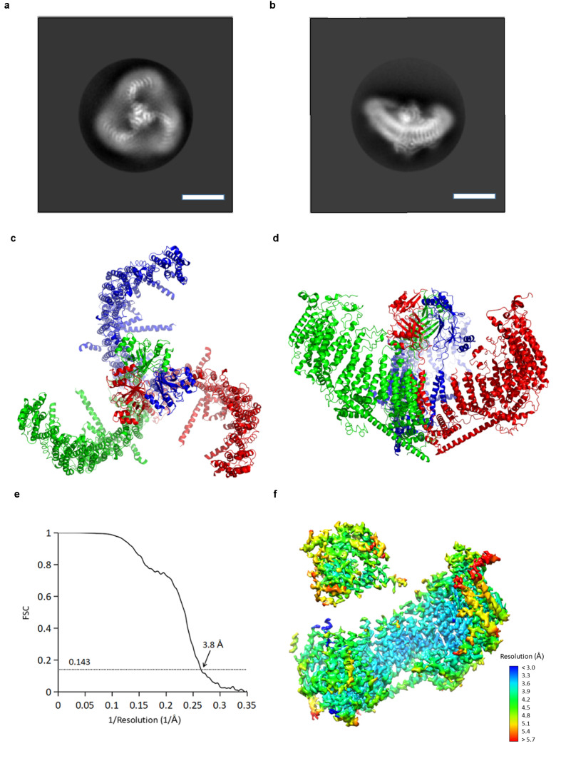
Cryo-EM structure of the Piezo1 mechanosensitive channel: the three-bladed propeller (2D class average, top) and the hom
the biology Cryo-EM structure of the Piezo1 mechanosensitive channel: the three-bladed propeller (2D class average, top) and the homotrimeric atomic model (three subunits, red/green/blue) — the tension-gated channel the model reads out.Guo & MacKinnon, eLife 2017;6:e33660 · CC BY 4.0

ECM, motility & measurementthe matrix, the crawl, and how quantities are read out

6-material ECM librarycollagen · fibrin · PA · HA · Matrigel · agarose — real Pa★ new
\(1\ \text{pN/µm}^2=1\ \text{Pa};\qquad E_\parallel/E_\perp=f(S)\ (S=\text{nematic order})\)

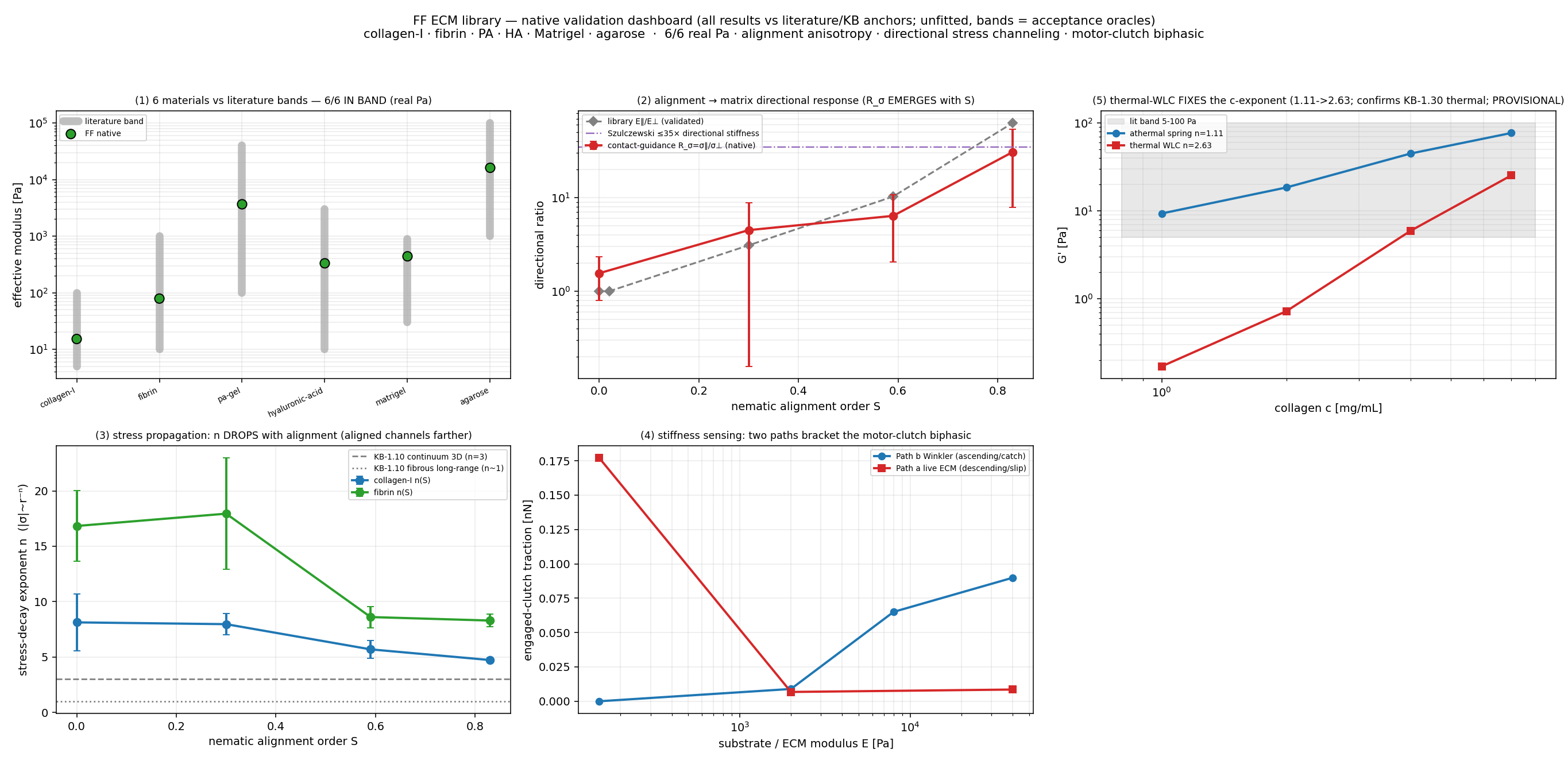
A grounded, alignment-controlled ECM library with two constitutive classes: fibrillar (collagen, fibrin) whose macroscopic modulus EMERGES from the Mikado microstructure, and continuum gels (PA/HA/Matrigel/agarose) as elastic spring lattices whose bond stiffness is the analytic inverse of the target E. A three-way harness reads shear G, uniaxial E (+anisotropy), and spherical indentation E_eff — all in SI Pascals (1 pN/µm² = 1 Pa exactly). Nematic order S about any director gives alignment→mechanical anisotropy; interpenetrating composites are supported.

Literature / anchor: Yang-Kaufman, Piechocka, Tse-Engler, Soofi, Normand (per-material bands); KB-1.10.

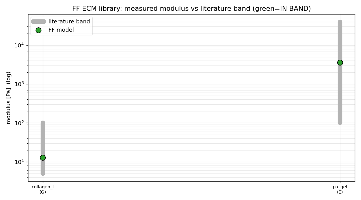
Validation: 6/6 materials land IN their literature band (collagen 12 Pa … agarose 14 kPa); alignment S=0.02→0.83 gives E∥/E⊥ 1→63× (loose stroma → tendon/scar); harness cross-validated on a known-modulus continuum.

All six ECM materials read out in real Pascals, each inside its literature band.
validation All six ECM materials read out in real Pascals, each inside its literature band.
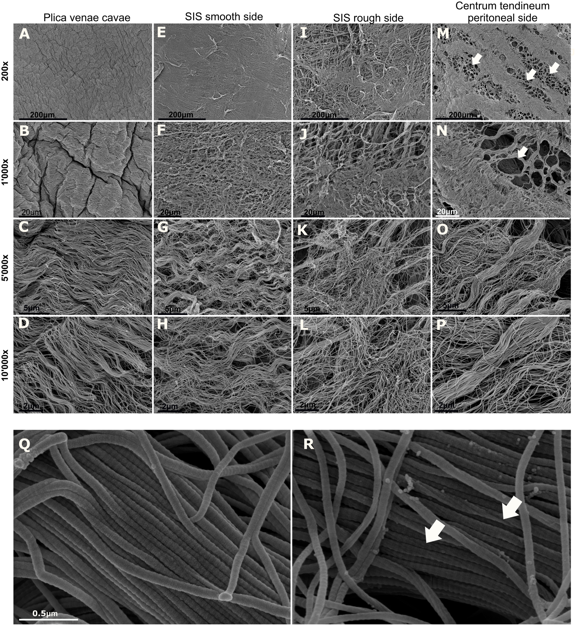
Scanning electron microscopy of a fibrillar collagen matrix at increasing magnification — the 3D collagen fiber meshwork
the biology Scanning electron microscopy of a fibrillar collagen matrix at increasing magnification — the 3D collagen fiber meshwork a cell grips and remodels (lower panels resolve individual fibrils with their ~67 nm D-banding).Maurer et al., PLoS ONE 2018;13:e0205027 · CC BY 4.0
ECM as 3D Mikado networkdisordered collagen rods, emergent mesh + stress decay● production path
\(\kappa=k_BT\,L_p,\qquad |\sigma(r)|\sim r^{-2}\ (\text{near-field dipole})\)

Disordered random straight collagen rods (uniform centre + isotropic orientation) discretized as Cytosim fibers, cross-linked by Hookean links at KDTree near-contacts, far boundary Dirichlet-pinned. Protrusion↔ECM via soft excluded-volume contact → load emergent; mesh size ξ is emergent (reported).

Literature / anchor: Wilhelm & Frey 2003; Head-Levine-MacKintosh 2003; collagen κ=k_BT·L_p (L_p=20µm).

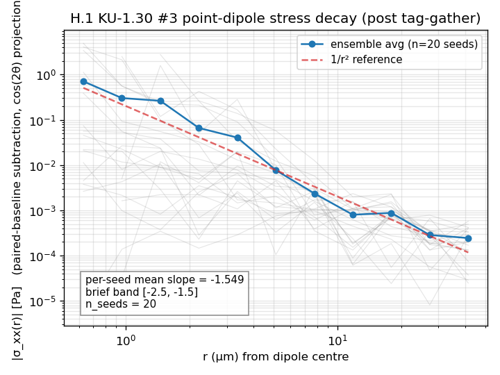
Validation: Reuses parity-checked fiber+link kernels; emergent ξ vs the Yang-Kaufman 1–5µm target; force-dipole stress decay near 1/r² in the network.

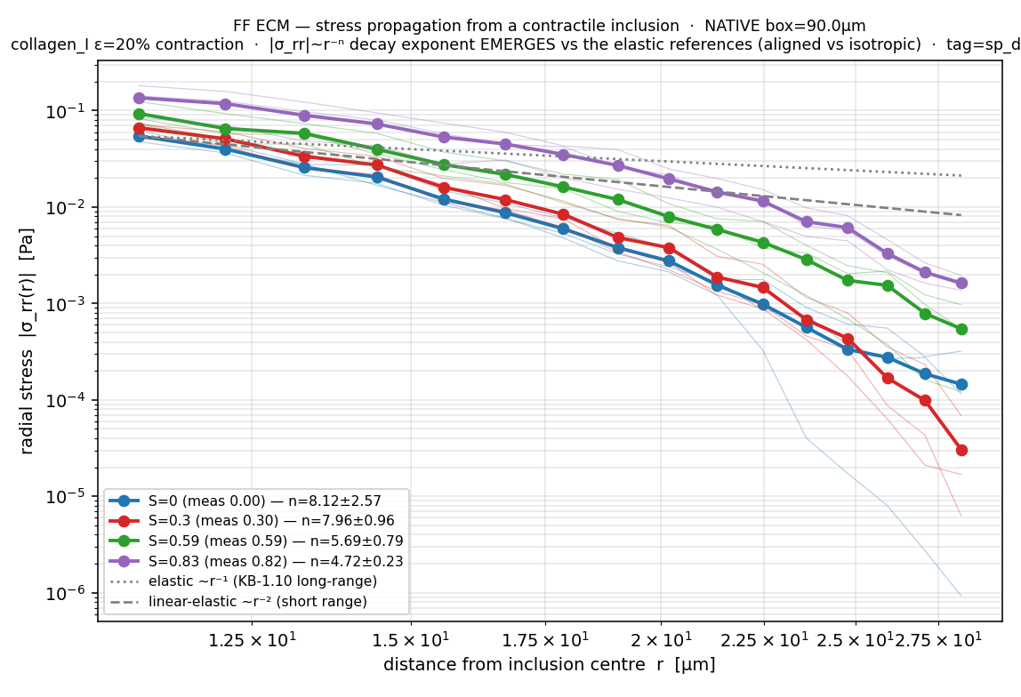
Directional stress propagation — an aligned collagen matrix channels a contractile inclusion's stress ~10× farther (nati
validation Directional stress propagation — an aligned collagen matrix channels a contractile inclusion's stress ~10× farther (native).
Scanning electron microscopy of a fibrillar collagen matrix at increasing magnification — the 3D collagen fiber meshwork
the biology Scanning electron microscopy of a fibrillar collagen matrix at increasing magnification — the 3D collagen fiber meshwork a cell grips and remodels (lower panels resolve individual fibrils with their ~67 nm D-banding).Maurer et al., PLoS ONE 2018;13:e0205027 · CC BY 4.0
Whole-cell crawl / motilityprotrusion + traction, physical-η closed loop△ honest shortfall
\(\dot{\mathbf x}_i=\dfrac{\Delta t}{\gamma_i}\,\mathbf F_i,\qquad \gamma_i\ \text{from}\ \eta=65.9\ \text{Pa·s}\)

Real overdamped step with per-node NF2007 cytoplasm drag (η=65.9 Pa·s) so time+speed are physical; leading-edge Mogilner-Oster protrusion (throttled by emergent counter-load) balanced by a uniform retrograde reaction (Newton's 3rd law); FA catch-slip clutches convert it to traction translocation. Adds spreading push, tension-gated area growth, gravity, volume feedback.

Literature / anchor: η=65.9 Pa·s Dessard 2024; KU-3.6 ratchet, KU-2.5 clutch, KU-3.12 retrograde flow.

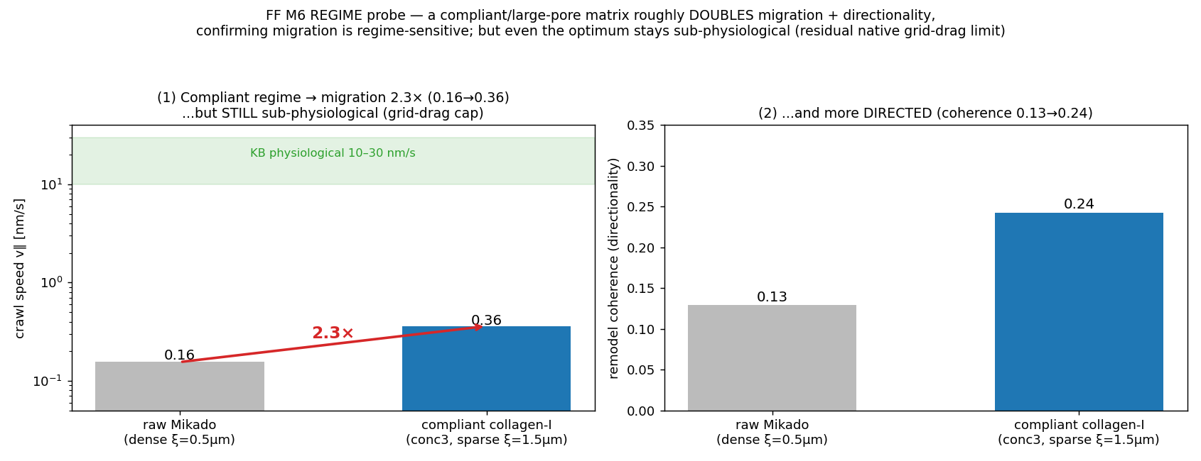
Validation: Without clutches the retrograde reaction makes net protrusion force = 0 (COM drift ≈ 0). Migration is regime-limited: matrix regime is a real ~2.3× lever (compliant large-pore collagen), while raw contractility/adhesion are NOT — a residual grid-drag/propulsion solver limit remains (open).

Migration lever probe — the ECM regime (pore size/compliance) is the real ~2.3× lever, not raw force.
validation Migration lever probe — the ECM regime (pore size/compliance) is the real ~2.3× lever, not raw force.
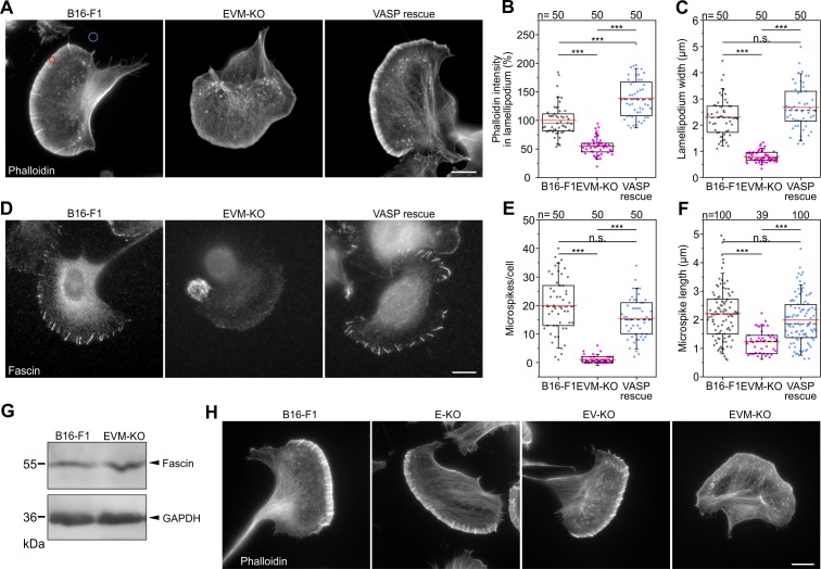
A B16-F1 cell migrating on laminin (F-actin, phalloidin) — a broad flat lamellipodium studded with filopodia at the lead
the biology A B16-F1 cell migrating on laminin (F-actin, phalloidin) — a broad flat lamellipodium studded with filopodia at the leading edge, the branched-actin protrusion machinery that drives crawling.Damiano-Guercio et al., eLife 2020;9:e55351 · CC BY 4.0
Cell polarizationactomyosin active-gel symmetry breaking✓ validated
\(\partial_t c=-\partial_x(cv)+D\,\partial_{xx}c-k_{\text{off}}(c-c_0)\)

A 1D periodic cortex-ring active gel: myosin advection-diffusion-turnover coupled to overdamped force balance with active stress σ_a=ζc/(1+c/c*); above the contractility threshold a single high-myosin cap (rear) forms with steady cortical flow → front-rear symmetry breaking. Solved spectrally.

Literature / anchor: Bois, Jülicher & Grill 2011 PRL 106:028103; Mayer 2010 (ℓ≈14µm).

Validation: Linear dispersion λ(k) measured-vs-analytic; single_cap_fraction ensemble honesty metric (robust single cap only near threshold — flagged after an audit caught seed cherry-pick).

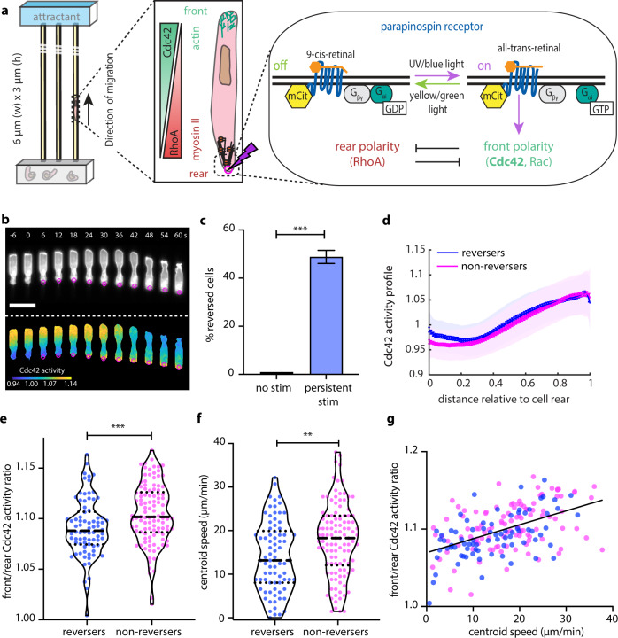
Front–rear polarity of a migrating cell: the leading edge (Cdc42–Rac, branched actin) vs the trailing rear/uropod (RhoA,
the biology Front–rear polarity of a migrating cell: the leading edge (Cdc42–Rac, branched actin) vs the trailing rear/uropod (RhoA, myosin II), with live Cdc42-activity concentrated at the front — actomyosin symmetry breaking that sets the migration axis.Hadjitheodorou et al., Nat. Commun. 2021;12:6619 · CC BY 4.0
Substrate plane + parallel-plate AFMthe measurement geometry, protocol-consistent● production path
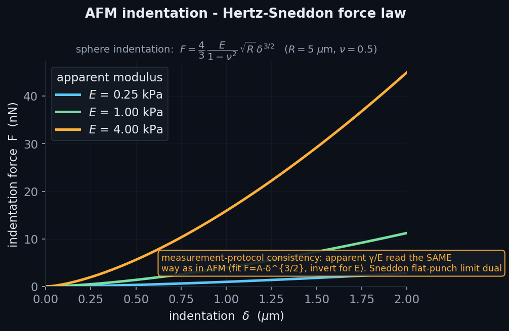
\(F=\tfrac{4}{3}\dfrac{E}{1-\nu^2}\sqrt{R}\,\delta^{3/2}\quad(\text{Hertz–Sneddon})\)

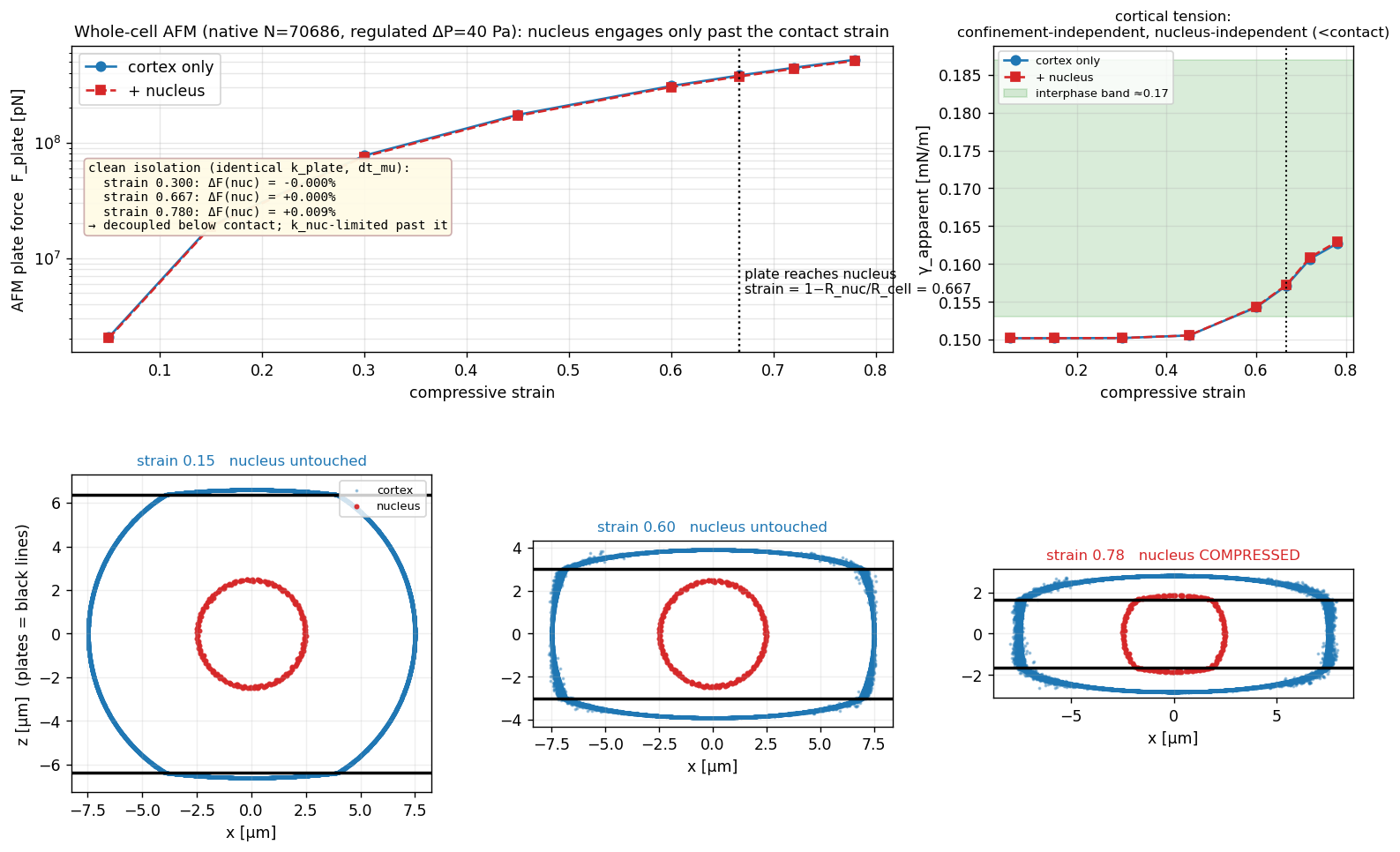
A one-sided z-penalty holds sub-plane nodes on the substrate; two rigid parallel plates compress the turgor-pressurised cortex and read the top-plate reaction = AFM force, at an η-limited physical press-speed ramp. The apparent γ is extracted the SAME way as experiment (liquid-drop Young-Laplace).

Literature / anchor: Fischer-Friedrich parallel-plate protocol; measurement-protocol-consistency sanity gate.

Validation: Rigid clamp is exact; press-speed ramp fixes the fast-press non-equilibrium transient.

Analytic Hertz–Sneddon indentation F∝E·δ^{3/2} — apparent modulus read the same way as in AFM (protocol consistency).
the physics Analytic Hertz–Sneddon indentation F∝E·δ^{3/2} — apparent modulus read the same way as in AFM (protocol consistency).
Traction-force microscopy of a single adherent cell: the substrate bead-displacement field and reconstructed traction-st
the biology Traction-force microscopy of a single adherent cell: the substrate bead-displacement field and reconstructed traction-stress map (concentrated at the adhesive corners) — the forces a cell transmits to its elastic substrate.Ruppel et al., PLoS Comput. Biol. 2026;22:e1014045 · CC BY 4.0
Virial stress + areal strainthe Cauchy stress field driving the viewers● production path
\(\boldsymbol\sigma_{\text{node}}=\dfrac{1}{V}\sum \tfrac12(\mathbf r_{ij}\otimes\mathbf f_{ij})-\Delta P\,\mathbf I\)

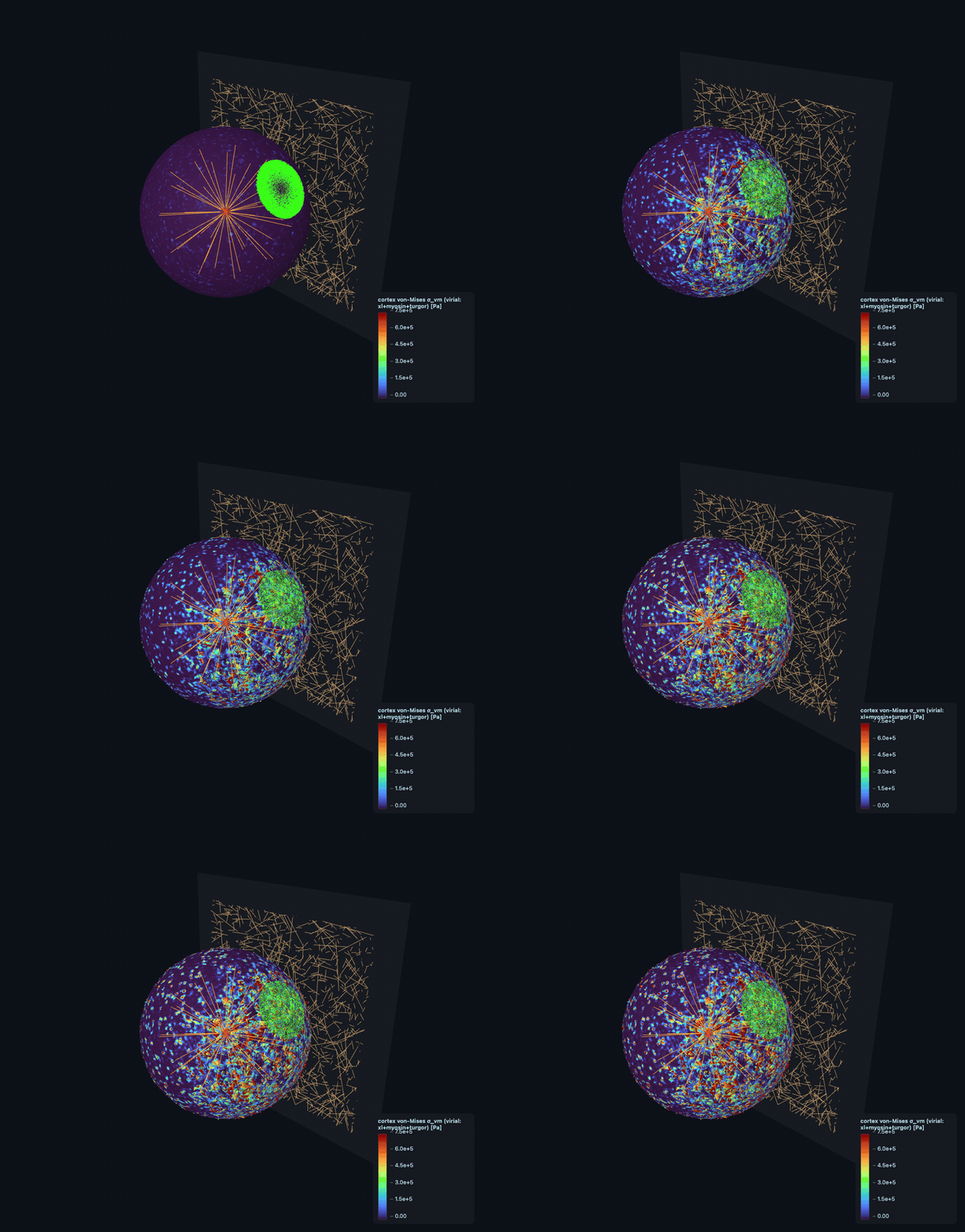
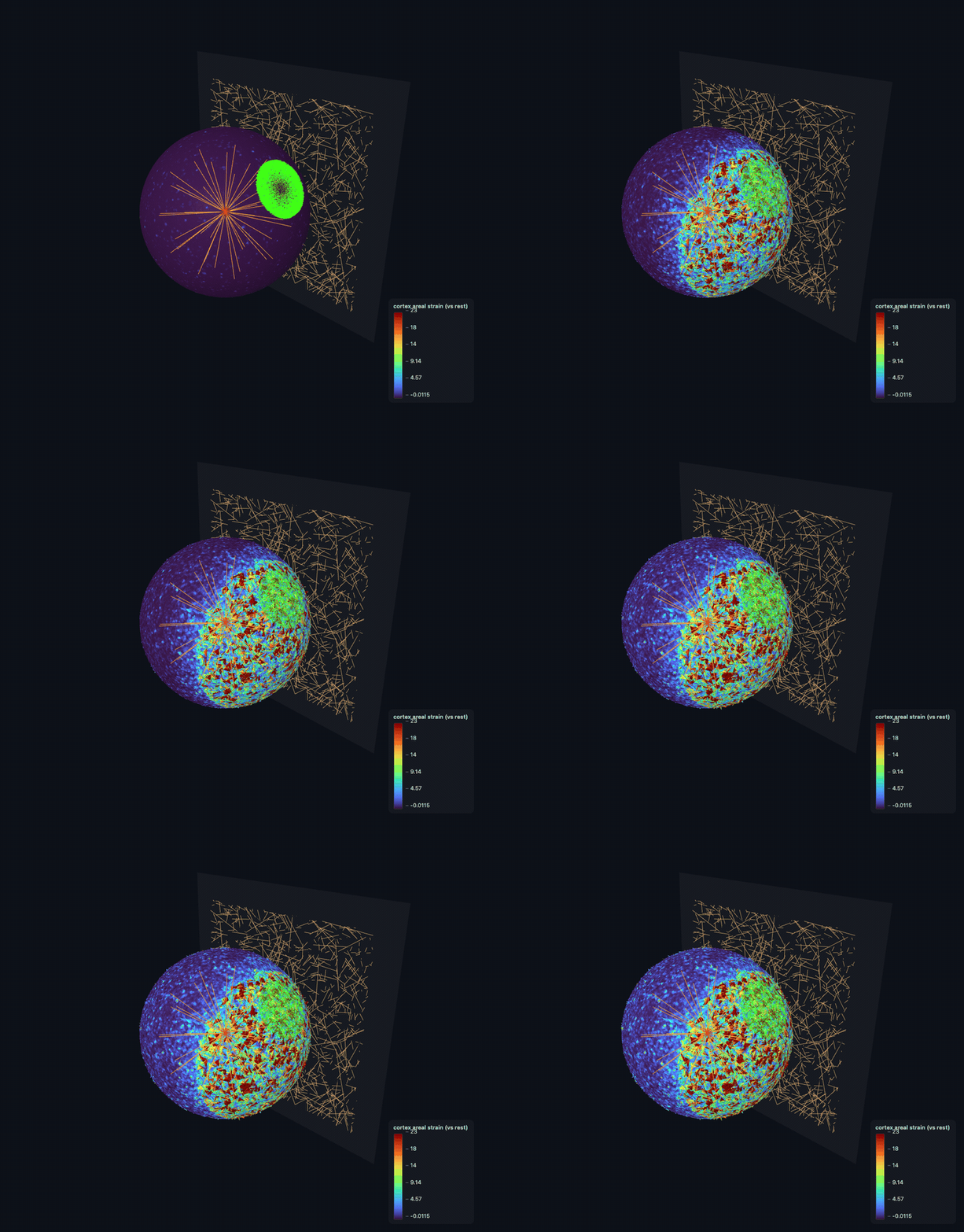
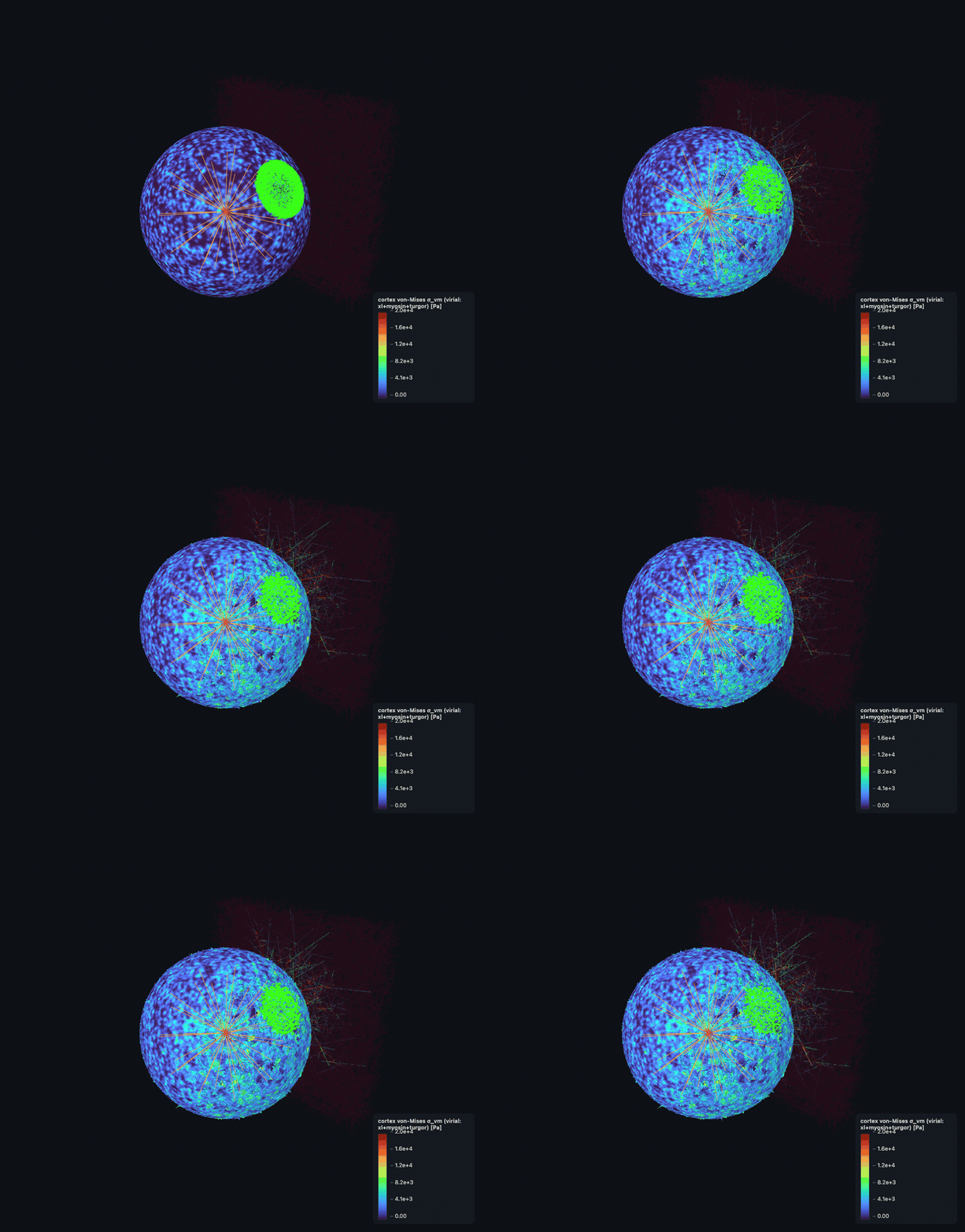
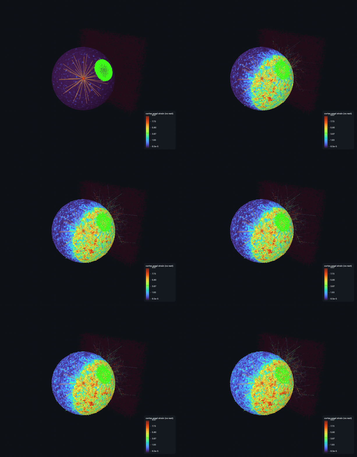
Per-node von-Mises stress from the virial tensor σ=(1/V)Σ½(r⊗f) over crosslink + myosin bonds plus the −ΔP·I turgor hydrostatic — a REAL internal stress (nonzero at equilibrium), not the net residual. Plus per-node areal strain from the fixed surface triangulation.

Literature / anchor: Cauchy/virial construction; pN/µm² = Pa exactly.

Validation: Diagnostic field for the flagship viewers (bending, a 3-body term, is excluded and the field is labelled accordingly).

Overdamped integratorexplicit CFL + NF2007 linearly-implicit solve● production path
\(\Big(\dfrac{\gamma}{\Delta t}I+K\Big)\Delta\mathbf x=\mathbf F,\qquad K=\alpha D^{\mathsf T}D+\textstyle\sum k\,\hat{\mathbf u}\hat{\mathbf u}^{\mathsf T}\)

Explicit projected overdamped descent x+=(dt/γ)·P·F (CFL dt<γ·seg³/8κ); and the linearly-implicit NF2007 step (γ/dt·I+K)·dx=F with K assembled ANALYTICALLY (bending 4th-diff + crosslink rank-1), LU-prefactored once or CG (cupy GPU-resident). Unconditionally stable → dt bounded by accuracy. A FF-specific solver was needed because the DCM FD-Jacobian CG blows up at FF force scales (~1e5–1e6 pN).

Literature / anchor: Nédélec & Foethke 2007 §5.2/Eq2 (10⁴×-class speedup).

Validation: Stiff bent fiber reaches few-% bending energy in 1–3 implicit steps; matches projected-explicit equilibrium to ~1e-6 µm.

DCM — Deformable Cell Model engine · the mesoscale whole-cell shell & multicellular tissue (SimuCell3D physics)

Cell shell & turgorthe deformable body: a pressurised elastic shell

Deformable triangulated surfaceclosed icosphere, nodes are the DOF● production path
\(V=\tfrac16\sum \mathbf v_0\cdot(\mathbf v_1\times\mathbf v_2)\quad(\text{divergence theorem})\)

Each cell is a closed outward-wound icosphere (42 or 162 nodes) of triangle faces; nodes are the integrated (overdamped) DOF. Faces carry turgor/contact/tension, edges carry cortical springs. Enclosed volume via the divergence theorem. A fixed-size node-pool keeps device topology static across division/remesh.

Literature / anchor: SimuCell3D (Runser/Vetter/Iber 2024); CellSim3D (Madhikar 2018).

Validation: Enclosed-volume + closed-manifold (Euler, outward-winding) invariants checked in-op by every remesh op.

Turgor (volume-conserving)exact-volume osmotic cytoplasm pressure● production path
\(\Delta P_c=\Delta P_0+K_{\text{vol}}\dfrac{V_0-V_c}{V_0},\qquad \mathbf F=\Delta P\,(A\,\hat{\mathbf n})\)

Two-pass exact-volume turgor: reduce per-cell V, then a linear osmotic ΔP=ΔP₀+K_vol·(V₀−V)/V₀ applied OUTWARD as F=ΔP·(area·n̂) split to the 3 face nodes. An osmotic variant scales ΔP₀·V₀/V for concentration feedback. ΔP₀=133 Pa MCF7 Young-Laplace baseline.

Literature / anchor: SimuCell3D compressible-fluid cytoplasm; MCF7 ΔP₀ Young-Laplace.

Validation: Force-law parity to machine-eps vs the committed fixture; analytic Laplace ΔP·R/2 ground truth.

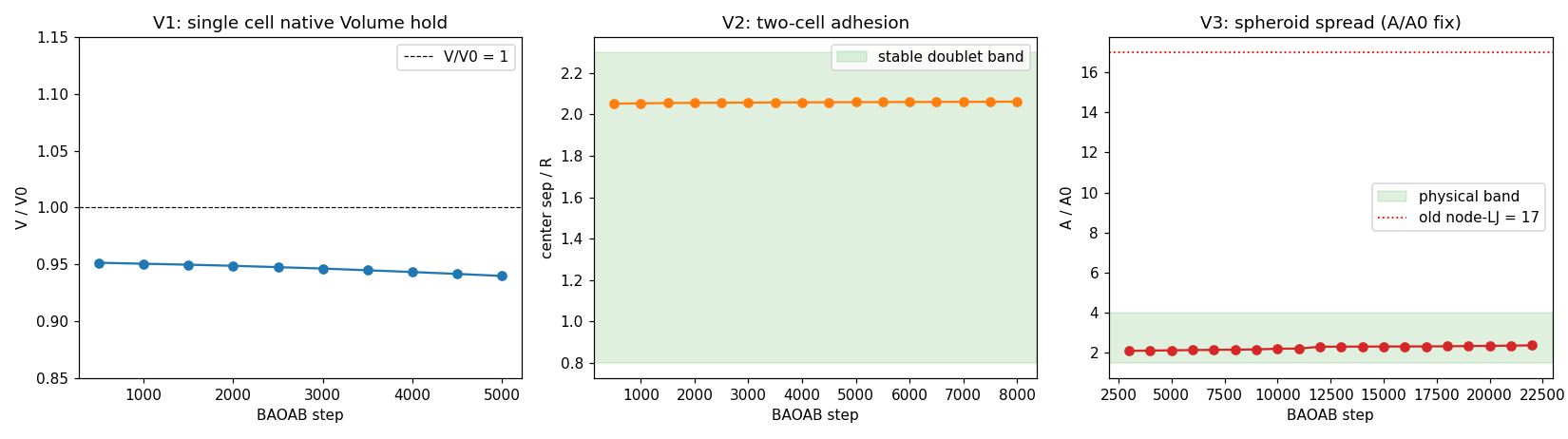
Single-cell volume conservation V/V₀ under turgor over a BAOAB run (honest ~6% drift, annotated).
validation Single-cell volume conservation V/V₀ under turgor over a BAOAB run (honest ~6% drift, annotated).
Cortical tension γsurface-energy area-minimising tension⚠ additive / PI-gated
\(\mathbf F=-\gamma\,\dfrac{\partial A}{\partial\mathbf r}\quad(\text{area-minimising, }\tilde\gamma=\gamma/K\ell)\)

surface_tension_kernel applies F=−γ·∂A/∂r per face scattered to its 3 vertices (analytic area gradients); an optional global-area force adds areal incompressibility. Cortical elasticity also enters as harmonic edge springs. Uniform γ alone leaves a rounded 'bag of marbles' — faceting needs differential tension + confluent init.

Literature / anchor: SimuCell3D shell surface-tension energy; nondim γ̃=γ/(K·l).

Validation: Area-gradient is analytic and momentum-clean by construction; nondim.py maps FF-measured γ → DCM γ̃.

Fluorescence of a cell — F-actin (green), the focal-adhesion marker paxillin (magenta), DNA (blue): the contractile acto
the biology Fluorescence of a cell — F-actin (green), the focal-adhesion marker paxillin (magenta), DNA (blue): the contractile actomyosin stress-fiber array that non-muscle myosin II assembles and tensions, anchored at focal adhesions.Weißenbruch et al., eLife 2021;10:e71888 · CC BY 4.0

Cohesion, contact & tissuecell–cell junctions, non-interpenetration, faceting, compaction

Cadherin catch-bond cohesionexplicit E-cadherin trans-dimer ensemble✓ validated
\(\mathbf F=k_{\text{trans}}\max(0,L-r_0);\quad k_{\text{off}}(F)\ \text{Rakshit},\ f_0\!\approx\!29\ \text{pN}\)

One attractive-only harmonic bond per apposed membrane-node pair of DIFFERENT cells (k_trans=5.84e-5 N/m); BREAK uses the force-dependent Rakshit sliding-rebinding catch-slip off-rate (exact 3×3 generator), FORM is mutual-nearest within r_bind. De-cohesion EMERGENT (bonds load past the catch peak f₀≈29pN into slip). GPU-native break/form kernels. Anti-lumped: replaced the binary cad_mult latch per PI.

Literature / anchor: Rakshit 2012 sliding-rebinding (KU-4.2); Iturri F_detach 6.5nN; k_on=27.96/s.

Validation: effective k_off from the exact generator; host↔GPU parity; KMC survival sub-cycled so large mechanics dt doesn't overshoot turnover.

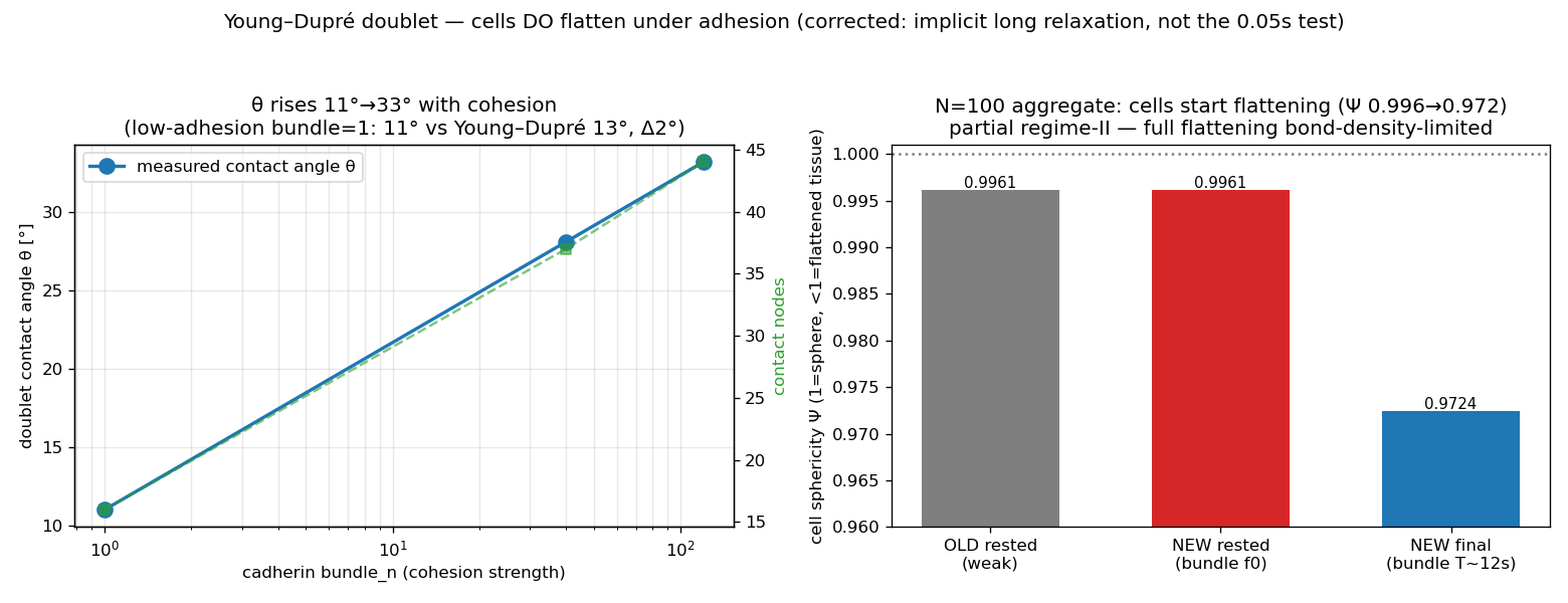
Cell-cell doublet contact angle vs cadherin cohesion — cells flatten along the Young-Dupré curve.
validation Cell-cell doublet contact angle vs cadherin cohesion — cells flatten along the Young-Dupré curve.
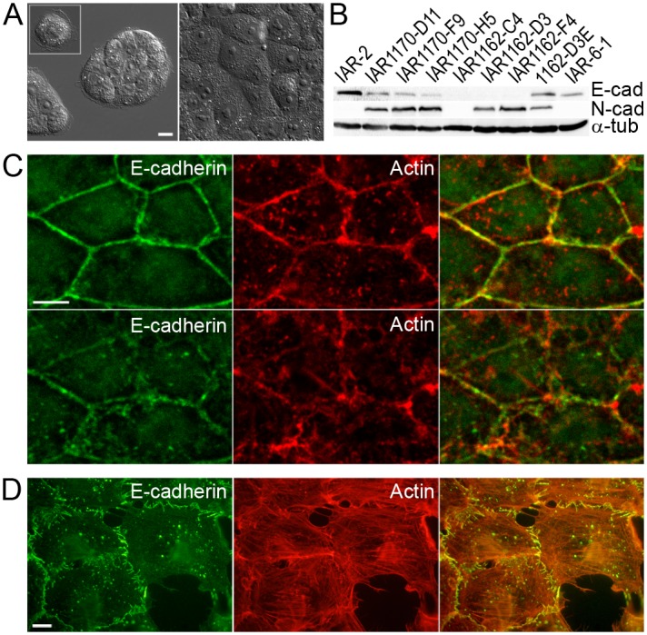
Confocal immunofluorescence of an epithelial sheet: E-cadherin (green) forms continuous adhesion belts tracing every cel
the biology Confocal immunofluorescence of an epithelial sheet: E-cadherin (green) forms continuous adhesion belts tracing every cell–cell border and co-localizing with the circumferential actin ring (red) — the adherens junction that holds neighbouring cells together.Rubtsova et al., PLoS ONE 2015;10:e0133578 · CC BY 4.0
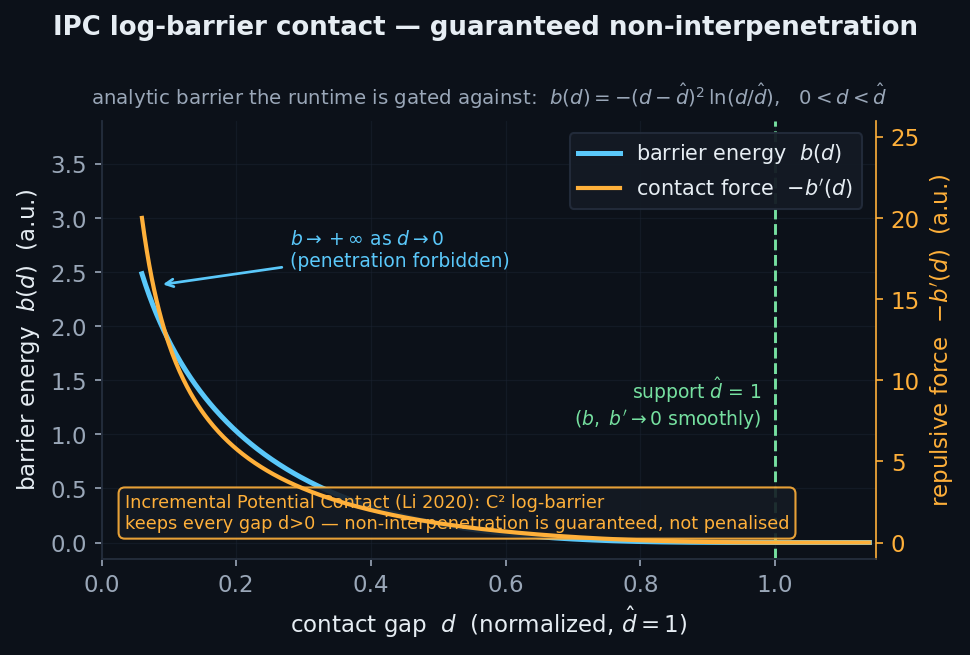
IPC log-barrier contact + CCDguaranteed non-interpenetration● production path
\(b(d)=-(d-\hat d)^2\ln(d/\hat d)\ \ [0

Node-vs-nearest-other-cell-face IPC barrier b(d)=−(d−d̂)²ln(d/d̂) with analytic derivatives; contact stiffness fed to the matrix-free CG so the implicit solve ABSORBS contact; CCD conservative-advancement α-clamp guarantees a penetration-free step. Supersedes the plain penalty contact (which tunnels or diverges).

Literature / anchor: Incremental Potential Contact (Li et al. 2020); SimuCell3D node-face; Ericson closest-point.

Validation: In-module self-tests (barrier / CCD / projection) + per-frame max-penetration diagnostic; energy kernels mirror the force kernels term-by-term.

Analytic IPC log-barrier b(d)=−(d−d̂)²ln(d/d̂) → +∞ as d→0, guaranteeing non-interpenetration.
the physics Analytic IPC log-barrier b(d)=−(d−d̂)²ln(d/d̂) → +∞ as d→0, guaranteeing non-interpenetration.
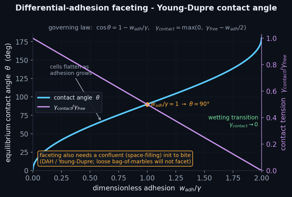
Differential-adhesion facetingDAH/foam interfacial tension → space-filling⚠ additive / PI-gated
\(\cos\theta=1-\dfrac{w_{\text{adh}}}{\gamma},\qquad \gamma_{\text{contact}}=\max(0,\gamma_{\text{free}}-\tfrac{w_{\text{adh}}}{2})\)

A cell-cell CONTACT face gets REDUCED tension γ_contact=max(0,γ_free−w_adh/2); free faces keep γ_free. The system lowers energy by growing contact area (Young-Dupré) → a space-filling faceted foam. An apico-basal polarized variant wets basal faces. Faceting was diagnosed as a confluent-init problem: gapped icospheres can't assemble into facets.

Literature / anchor: Steinberg DAH; Manning PNAS 2010; Douezan spreading S=w_cs−2γ; w_cs=2.85e-3 J/m².

Validation: Regime SET by literature values (no outcome tuning); reuses the validated area-gradient + face-contact detection.

Analytic Young–Dupré contact angle cosθ=1−w_adh/γ — cells flatten as adhesion grows (faceting also needs confluent init)
the physics Analytic Young–Dupré contact angle cosθ=1−w_adh/γ — cells flatten as adhesion grows (faceting also needs confluent init).
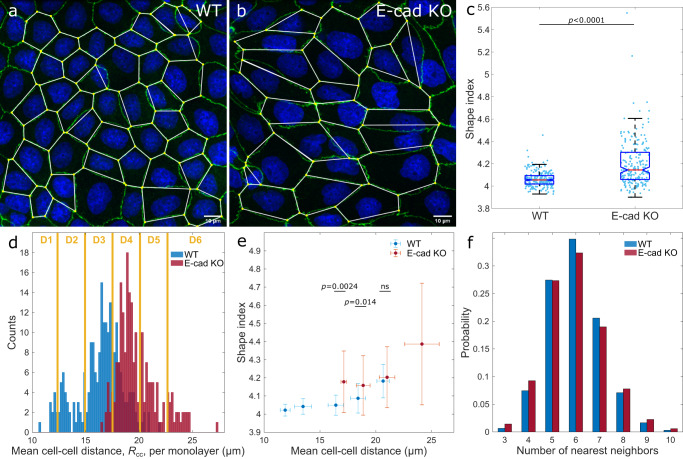
A confluent MDCK epithelial monolayer stained for the junction protein ZO-1 (green): apical cell–cell junctions form a f
the biology A confluent MDCK epithelial monolayer stained for the junction protein ZO-1 (green): apical cell–cell junctions form a foam-like polygonal (predominantly hexagonal) tessellation — the tissue's cell-packing topology, with segmented cell shapes overlaid.Eckert et al., Nat. Commun. 2023;14:5762 · CC BY 4.0
Aggregate tissue surface tensionwhole-spheroid Laplace → compaction⚠ additive / PI-gated
\(\Delta P_{\text{agg}}=\dfrac{2\sigma}{R_{\text{agg}}}\quad(\text{inward, media-exposed faces})\)

The whole spheroid is treated as a liquid drop of surface tension σ: Laplace ΔP_agg=2σ/R_agg applied INWARD (toward the global centroid) on media-exposed faces only, area-weighted. It globally densifies (void elimination) at constant cell volume — what per-cell/pairwise γ cannot do.

Literature / anchor: Foty-Steinberg tissue surface tension (~1–20 mN/m band).

Validation: Global centroid-radius reduction (unit-consistent); σ swept over the literature band, not tuned to a compaction target. Stable only on confluent init.

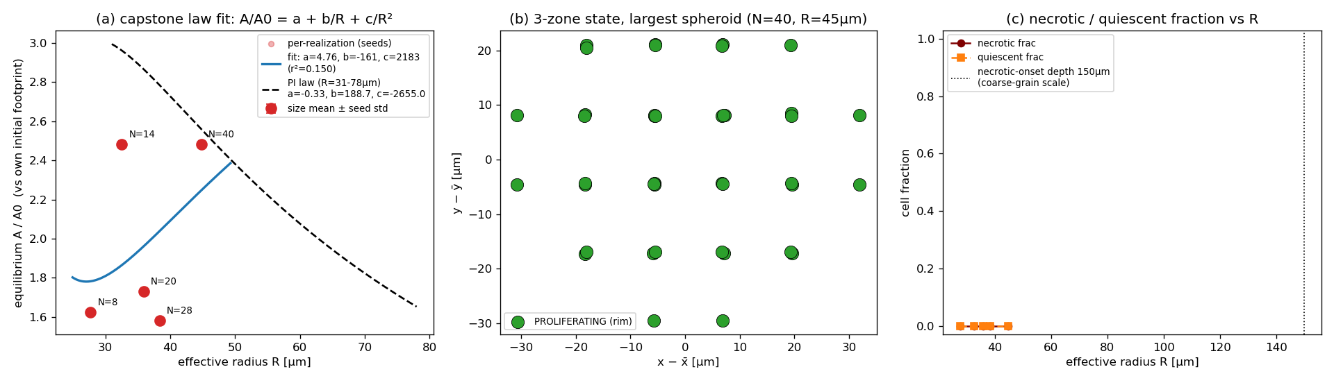
DCM equilibrium A/A₀ vs spheroid radius R — recovers the a+b/R+c/R² spreading FORM (magnitude a structural limit, honest
validation DCM equilibrium A/A₀ vs spheroid radius R — recovers the a+b/R+c/R² spreading FORM (magnitude a structural limit, honest).
Confocal cross-sections of tumour spheroids (FUCCI cell-cycle labelling) — the canonical concentric 3D architecture: an
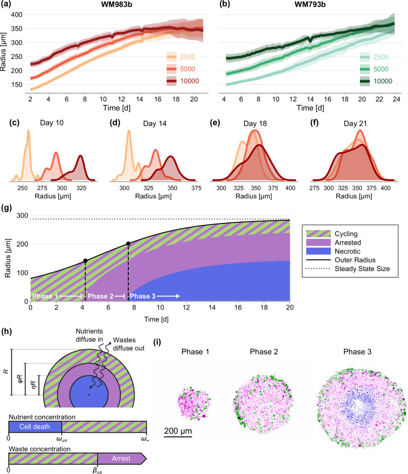
the biology Confocal cross-sections of tumour spheroids (FUCCI cell-cycle labelling) — the canonical concentric 3D architecture: an outer rim of cycling cells (green), an arrested zone (magenta) and a central necrotic core.Browning et al., eLife 2021;10:e73020 · CC BY 4.0
T1 rate-driven rearrangementevent-driven unjamming → biology-time tissue flow★ new
\(P(\text{T1})=1-e^{-k_{T1}\Delta t}\ \Rightarrow\ s\to s_0^{*}\ (\text{unjam})\)

A kinetic-Monte-Carlo T1 neighbour-exchange move at a physiological rate k_T1 supplies the sub-dt topological rearrangements that the large-dt mechanics correctly skips. It fluidizes a jammed confluent tissue so a spheroid can flow and remodel over BIOLOGY time (~600 s) instead of freezing — reaching the same unjammed state ~20× faster in wall-clock. The mechanics still freeze; the rate-T1 supplies the flow.

Literature / anchor: Kim τ_p=2^N·τ compaction-rate framework; SPV/vertex-model T1 unjamming.

Validation: 12-test KMC suite + k_T1(s) calibration B=2.68; the biology-time HERO run fluidizes (s→5.02 unjam, 3.29µm rearrangement, A/A₀ 1.04) vs the frozen jam (s4.94, disp 0.08). Honest: the rigid-move step is not strictly shape-neutral (refinement).

A T1 (cell-neighbour-exchange) transition in a vertex model of an epithelium: a shrinking junction (red) collapses to a
the biology A T1 (cell-neighbour-exchange) transition in a vertex model of an epithelium: a shrinking junction (red) collapses to a four-fold vertex and a new junction opens perpendicular (green), so two cells swap neighbours — the local rearrangement that fluidizes tissue.Sknepnek et al., eLife 2023;12:e79862 · CC BY 4.0

Compartments, division & measurementnucleus, division, ECM, remesh, integrator, stress

Nucleus compartmentsolid-core excluded-volume confinement● production path
\(k_{\text{chrom}}=\dfrac{4\pi E_{\text{nuc}} R_{\text{nuc}}}{n}\quad(\text{N-invariant bridge})\)

The nucleus is a solid deformable stiff core: a one-sided OUTWARD bilinear push felt when a membrane node intrudes within R_nuc of the cell centroid (soft chromatin up to the lamin knee, stiff lamin beyond); force-free at r=R_nuc, never pulls inward. k_chrom is an N-invariant continuum bridge.

Literature / anchor: H.9 KU-3.B2 (bilinear nucleus); E_nuc=3e3 Pa, ratio_lamin=3, knee_strain=0.10.

Validation: The parity-tested radial_shell law-0 is the symmetric-bilinear shell; this centroid-based one-sided kernel shares the continuum bridge (R_nuc=0.25 found ~3× undersized, surfaced).

3D-SIM super-resolution image of the nuclear lamina — the lamin meshwork resolves as a dense fibrous network enclosing g
the biology 3D-SIM super-resolution image of the nuclear lamina — the lamin meshwork resolves as a dense fibrous network enclosing gaps, the stiff strain-stiffening shell around the nucleus.Kittisopikul et al., Cells 2019;8:361 · CC BY 4.0
Membrane/cortex/cytoplasm stackthe shared 3-law radial-shell compartment● production path
\(\Delta P_{\text{mem}}=\dfrac{2\gamma}{R};\quad \Delta P_{\text{turgor}}=\Delta P_0-K_{\text{vol}}\dfrac{V-V_0}{V_0}\)

radial_shell is the shared 3-law compartment force over tagged shell beads: law-1 membrane Laplace ΔP=2γ/R (inward), law-2 turgor (outward), law-0 nucleus bilinear. In the multicell runtime these are realized per-cell as edge springs (cortex) + surface tension (membrane) + exact-volume turgor (cytoplasm) + nucleus core.

Literature / anchor: SimuCell3D; MembraneSurfaceTension / EnclosedVolumePressure / NucleusConfinement (HOOMD .cu).

Validation: Force-law parity ~1e-13 vs the numpy reference; mirrors the .cu term-by-term.

Cryo-electron tomography of an unroofed mammalian plasma membrane and the cortical actin meshwork beneath it (cyan = cor
the biology Cryo-electron tomography of an unroofed mammalian plasma membrane and the cortical actin meshwork beneath it (cyan = cortical filaments) — the membrane–cortex the model treats as an independent lipid sheet on the cortex.Sun et al., Nat. Commun. 2025;16:855 · CC BY 4.0
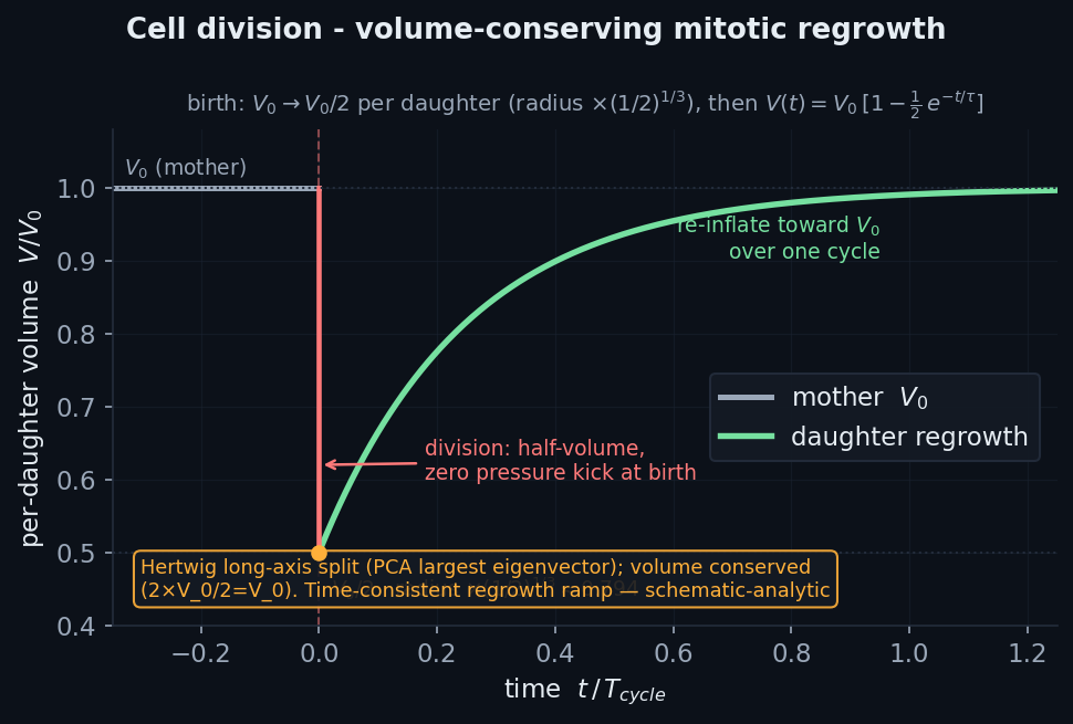
Proliferation / divisionvolume-conserving mitotic cytokinesis● production path
\(R\to R\,(\tfrac12)^{1/3},\qquad V_0\to V_0/2\ \ (\text{Hertwig long axis})\)

Rim cells divide at p_div per tick by mitotic-rounding volume-conserving cytokinesis — the mother splits into two half-volume icospheres offset along the Hertwig long axis (PCA largest eigenvector), each V₀ set to V₀/2 (zero pressure kick at birth), then a time-consistent regrowth ramp re-inflates over one cell cycle. In-place mesh cleave is also implemented.

Literature / anchor: CellSim3D division (Madhikar 2018); Hertwig long-axis rule; p_div=0.04.

Validation: Deterministic single-split test hook; mirrors the HOOMD ProliferationUpdater; the in-place cleave path has its own test.

Analytic volume-conserving mitotic cytokinesis: drop to V₀/2 (radius ×0.794), then time-consistent regrowth over one cyc
the physics Analytic volume-conserving mitotic cytokinesis: drop to V₀/2 (radius ×0.794), then time-consistent regrowth over one cycle.
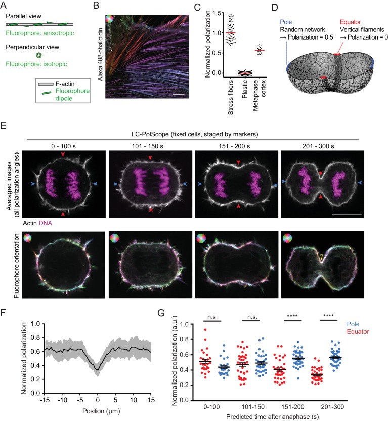
A dividing HeLa cell through cytokinesis (actin white, DNA magenta): the equatorial cleavage furrow ingresses as the con
the biology A dividing HeLa cell through cytokinesis (actin white, DNA magenta): the equatorial cleavage furrow ingresses as the contractile actomyosin ring constricts, pinching one cell into two daughters.Spira et al., eLife 2017;6:e30867 · CC BY 4.0
ECM / substrate couplingintegrin catch-slip clutch on a planar substrate● production path
\(k_{\text{off}}(F)=k_s\,e^{F/F_s}+k_c\,e^{-F/F_c}\quad(\text{Pereverzev})\)

Basal membrane nodes engage fixed substrate ligand sites as explicit integrin clutches (harmonic spring to a dish site); host BREAK at the Pereverzev two-pathway catch-slip off-rate, ENGAGE on-rate ∝ ligand density (Bare/Pre/Lam4). Plus a conservative substrate z-well and in-plane wetting. (Fiber ECM lives in the FF/collagen layer.)

Literature / anchor: Pereverzev two-pathway (KU-2.18); MCF7-on-pV4D4/col-I ligand ordering.

Validation: Pereverzev k_off from the same law as the FF clutch; substrate well/wetting parity to machine-eps.

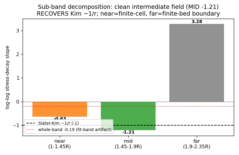
Fiber-ECM cell-induced strain/stress radial decay — recovers Kim ~1/r in the clean MID band (honest).
validation Fiber-ECM cell-induced strain/stress radial decay — recovers Kim ~1/r in the clean MID band (honest).
Actin stress fibers (magenta) terminating at focal adhesions marked by paxillin (green puncta), with a schematic of the
the biology Actin stress fibers (magenta) terminating at focal adhesions marked by paxillin (green puncta), with a schematic of the stress-fiber/adhesion organization — the molecular clutches that transmit traction to the ECM.Hauke et al., PLoS ONE 2021;16:e0250749 · CC BY 4.0
Adaptive remeshingedge-length + sliver-quality local refinement● production path
\(\ell\in[\ell_{\min},3\ell_{\min}],\qquad S_f=\dfrac{36A}{\sqrt3\,P^2}>0.2\)

Host-side low-cadence remesh keeps every edge in [l_min, 3·l_min] and no sliver face: priority SWAP → SPLIT (activate a dormant pool node) → COLLAPSE (park a node, non-manifold guard). Each op preserves the closed manifold + outward winding on a fixed-size pool (static device topology).

Literature / anchor: SimuCell3D local_mesh_refiner (sliver score from the source constexpr).

Validation: Enclosed-volume + Euler + boundary-manifold invariants checked each op; swap accepted only if quality strictly improves.

Overdamped integrator + IMEX NewtonBAOAB explicit + linearly-implicit contact solve● production path
\(\mathbf r(t{+}\Delta t)=\mathbf r+\dfrac{\mathbf F}{\gamma}\Delta t+\sqrt{\dfrac{k_BT}{2\gamma\Delta t}}(W_n{+}W_{n-1})\Delta t\)

Explicit path: overdamped Leimkuhler-Matthews BAOAB step. Implicit path: linearly-implicit Euler (γ/dt·I+K)Δx=F, matrix-free K·v via a device conjugate gradient; a projected-Newton IPC step re-linearises the stiff {turgor, edges, barrier} terms with Armijo line-search, plus a CCD α-clamp. kT=0 deterministic overdamped in production.

Literature / anchor: Leimkuhler-Matthews BAOAB-limit; implicit-Euler IMEX; per-node γ from η=65.9 Pa·s.

Validation: BAOAB bit-parity vs committed HOOMD fixtures; device-CG + IPC-Newton self-tests; autodiff vs finite-difference; M-SHAKE / Fixman vs fixtures.

Virial von-Mises stress fieldthe per-cell Cauchy stress the viewers colour by● production path
\(\sigma_{vm}=\sqrt{\tfrac32\,s_{ij}s_{ij}},\qquad p=-\tfrac13\operatorname{tr}\boldsymbol\sigma\)

node_virial_stress assembles the Cauchy/virial tensor over the dominant pairwise carriers — cadherin bonds + cortex edge springs (deviatoric) plus turgor isotropic −ΔP·I — and returns σ_vm and hydrostatic p per node in Pa. Built explicitly to FIX the viewer's old fmag channel (which was |residual force| ∝ velocity, not a stress).

Literature / anchor: Standard continuum virial/Cauchy stress; force laws match the sim kernels exactly.

Validation: Force laws reproduced term-by-term from the cadherin/edge/turgor kernels by construction; the net-force reconstruction is a self-consistency check.

Auxiliary EV + bending + osmoticedge-edge contact, Helfrich bending, sedimentation⚠ additive / PI-gated
\(\mathbf F_{\text{bend}}=-k_{\text{bend}}\,\Delta^2\mathbf r\quad(\text{Helfrich biharmonic})\)

edge-edge excluded volume (segment-segment) catches what node-vs-face misses; Helfrich-like thin-plate bending F=−k_bend·Δ²r resists curvature variation; a media-exposed fraction drives osmotic volume regulation; gravity adds sedimentation to the RHS. All additive/default-off.

Literature / anchor: Helfrich bending; KB-3.9 osmotic regulation; Ericson segment-segment.

Validation: Edge-edge is symmetric/momentum-clean; biharmonic discrete-Laplacian; gravity adds to the RHS only — all by construction.

Legacy lumped adhesiontent cohesion + binary junction switch⊘ superseded
\(\mathbf F=-\omega A\,m\,\text{tent}(r)+\text{rep},\qquad m=\sqrt{cad_1\,cad_2}\)

A bilinear node-node adhesion tent + repulsion, and a crowd-pressure-switched binary cad-multiplier junction latch. Explicitly VIOLATES the mechanistic hard rule and is SUPERSEDED by the explicit Rakshit cadherin catch-bond ensemble (PI 2026-06-21). Retained only as a parity-gated building block; production de-cohesion runs use cadherin bonds with these OFF.

Literature / anchor: DcmTentContact (SimuCell3D-style); slip-only cadherin KU-4.17.

Validation: Parity vs the HOOMD tent/cohesion fixtures; grid kernel reproduces brute-force to summation-order tolerance.

Play with a cell

Grab a fiber-network cell with your mouse and stretch it — a real-time toy (position-based dynamics on a real coarse FF cortex; not the fine-grained engine) for feeling how cells deform and spring back.

🖐 Grab & stretch — interactive cell

Real coarse FF actin cortex (up to ~31k nodes) + a translucent membrane and blue nucleus, driven by an adaptive real-time solver. Fibers colour by strain (blue = squished, red = stretched). Drag empty space to rotate.

▶ Open the playground

Cell morphology — flagship viewers & snapshots

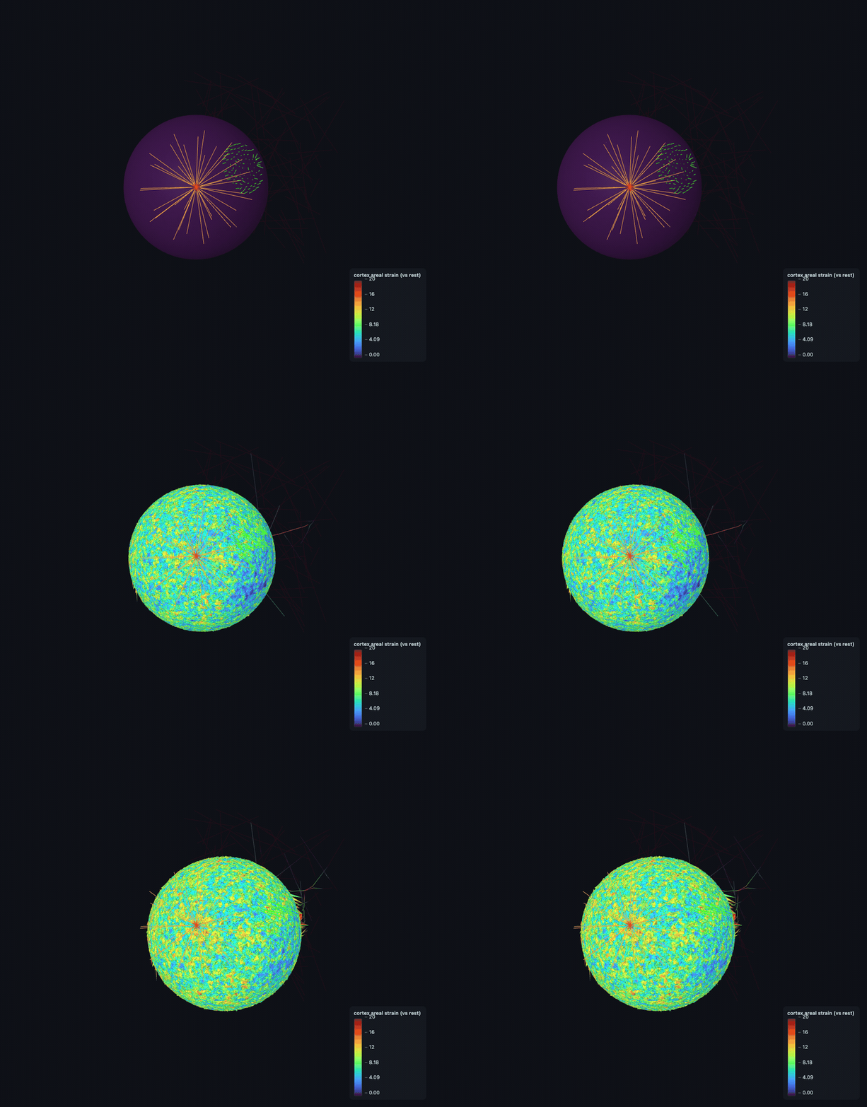
The heart of the project: interactive 3D cell morphology from each engine. The native-resolution FF flagships are 200–500 MB (too large for this site) — their real-viewer animations (the viewer's own shape / von-Mises stress / areal-strain scenes) are shown below and the full interactive HTML is a download link; the DCM flagship and the others open live here.

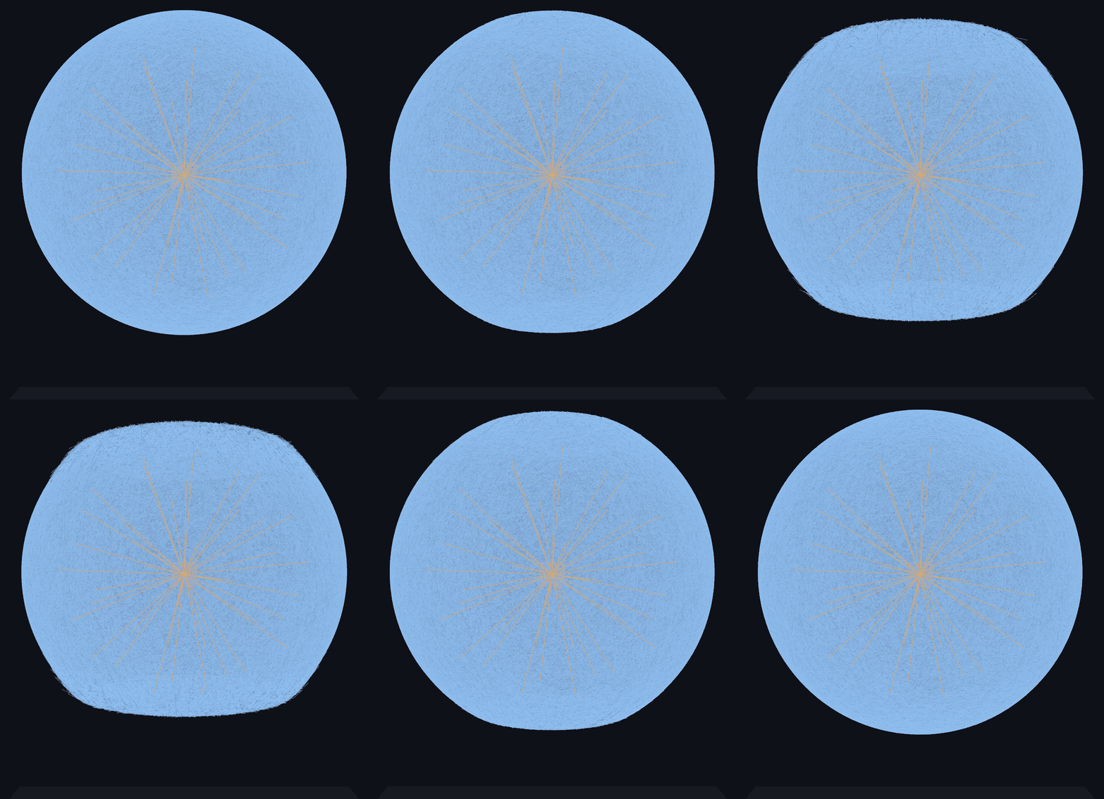
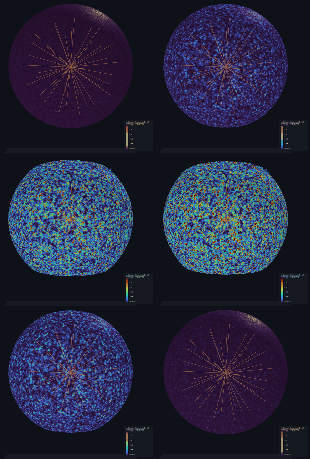
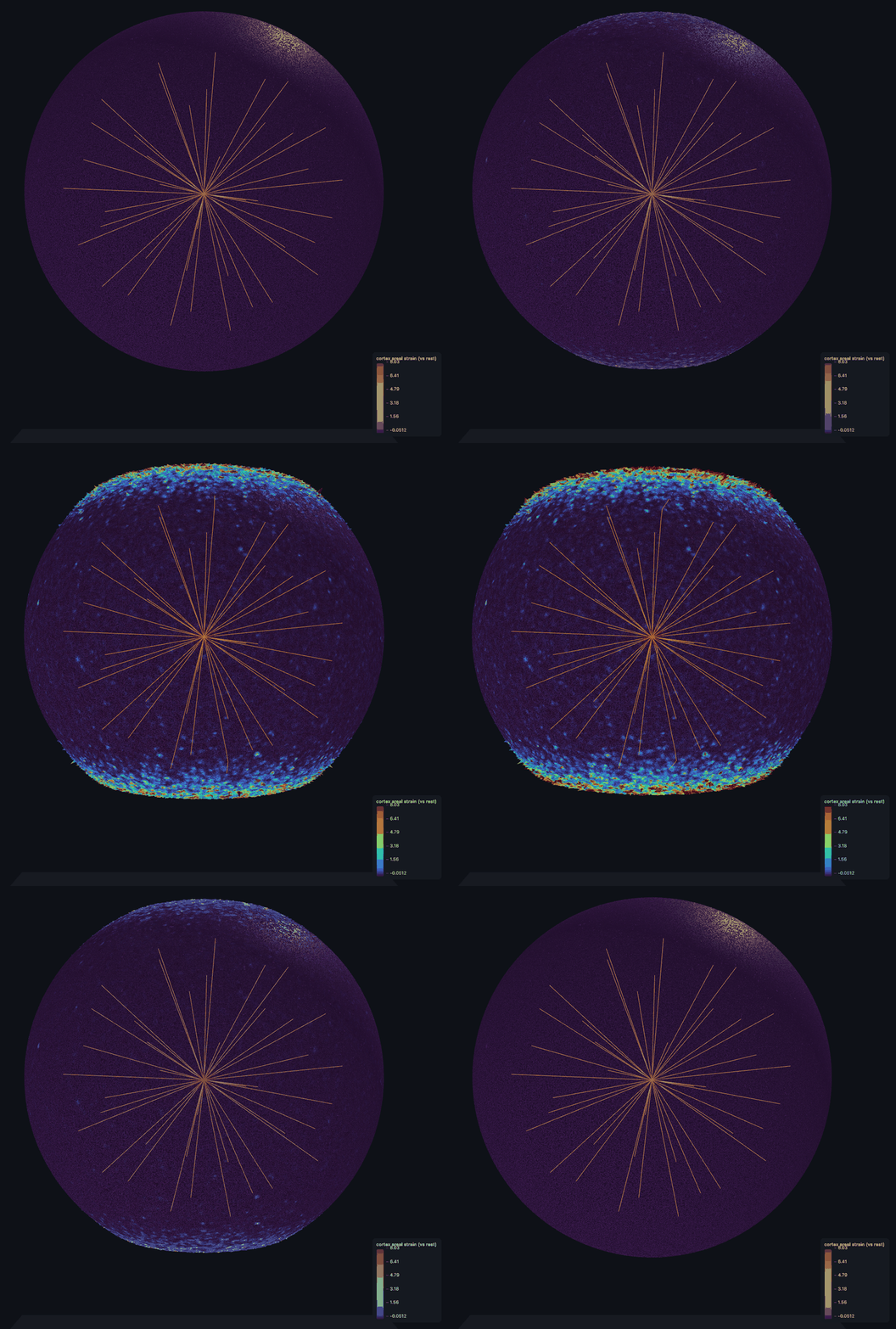
FF — AFM full-compartment indentation (native resolution)

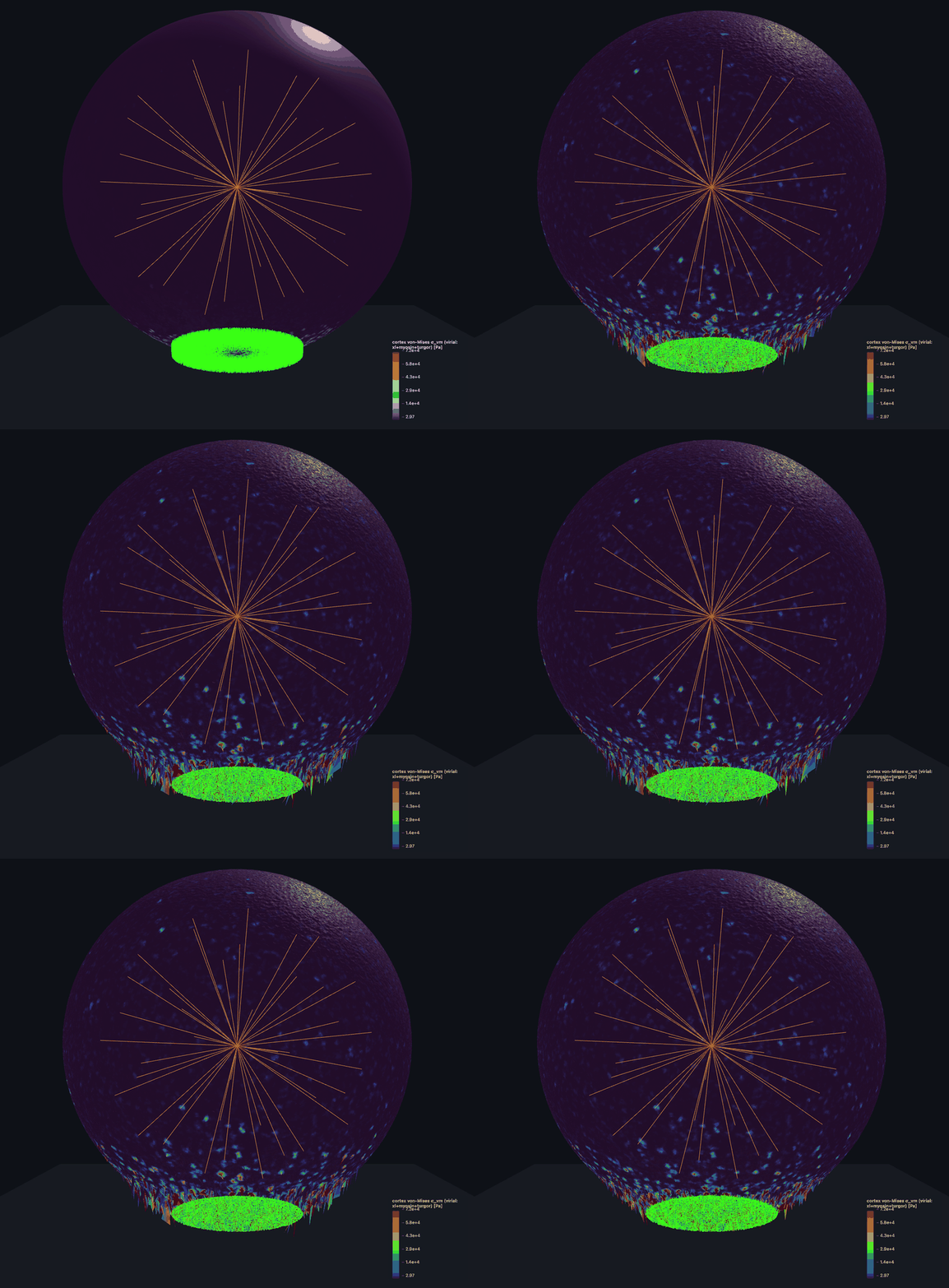
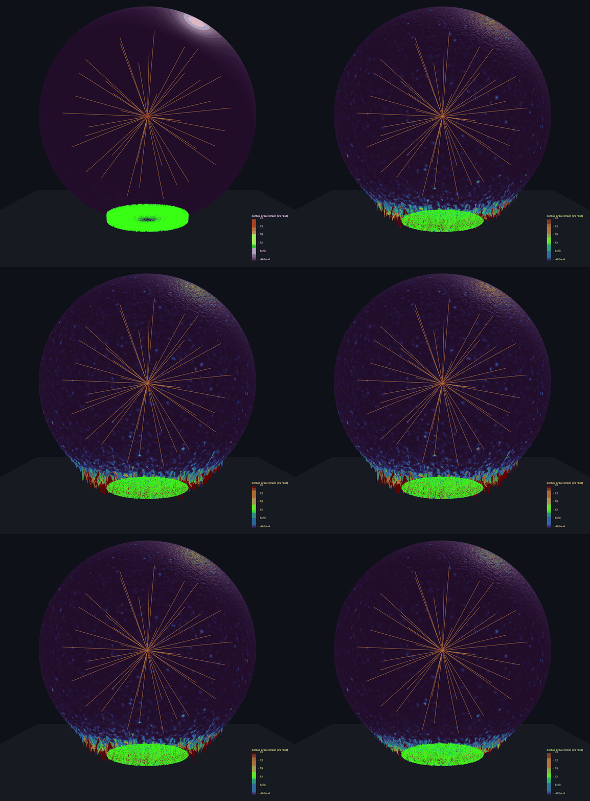
Woven-actin cortex + hull + microtubule aster (MTOC) + nucleus, indented by a rigid AFM plate. The animation is the real viewer's own morph (front view) — the cell visibly flattens and spreads; the two stills are its von-Mises stress and areal-strain fields.
indentation morph — the cell flattens &amp; spreads under the AFM plate — shape
▶ shape — indentation morph — the cell flattens &amp; spreads under the AFM plate
von-Mises stress morph
▶ σ_vm von-Mises stress (animated)
areal strain morph
▶ areal strain (animated)

⬇ Download full interactive viewer (500 MB)

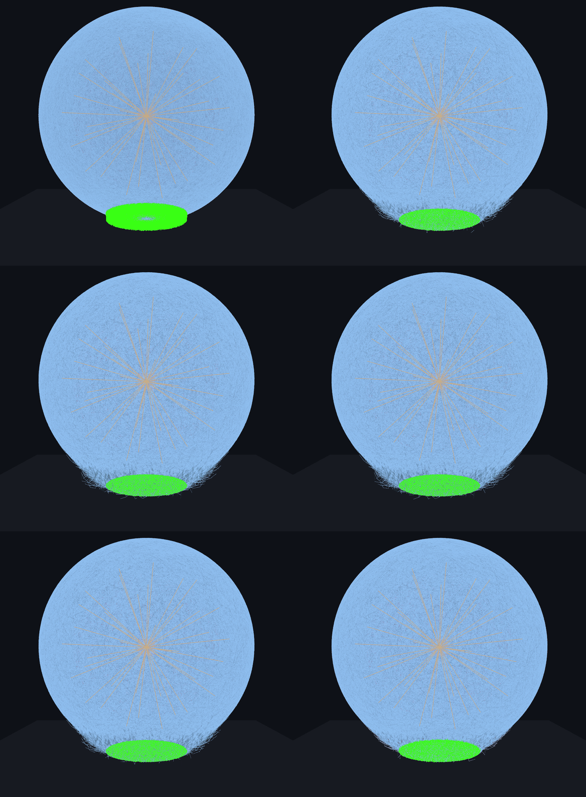
FF — microtubule + native cortex, cut (native resolution)

Native cortex with the microtubule aster and a green FA integrin-clutch patch on the substrate. The animation is the real viewer's morph (front view) settling onto the substrate; the two stills are its stress and strain fields.
indentation morph — cortex + MT aster settling onto the FA contact patch — shape
▶ shape — indentation morph — cortex + MT aster settling onto the FA contact patch
von-Mises stress morph
▶ σ_vm von-Mises stress (animated)
areal strain morph
▶ areal strain (animated)

⬇ Download full interactive viewer (509 MB)

FF — MCF7 cell remodels a collagen-I ECM (native resolution)

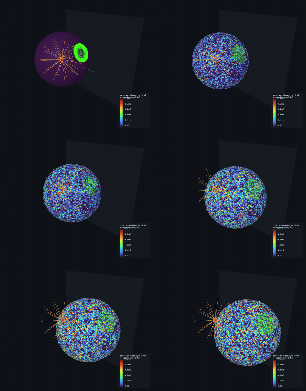
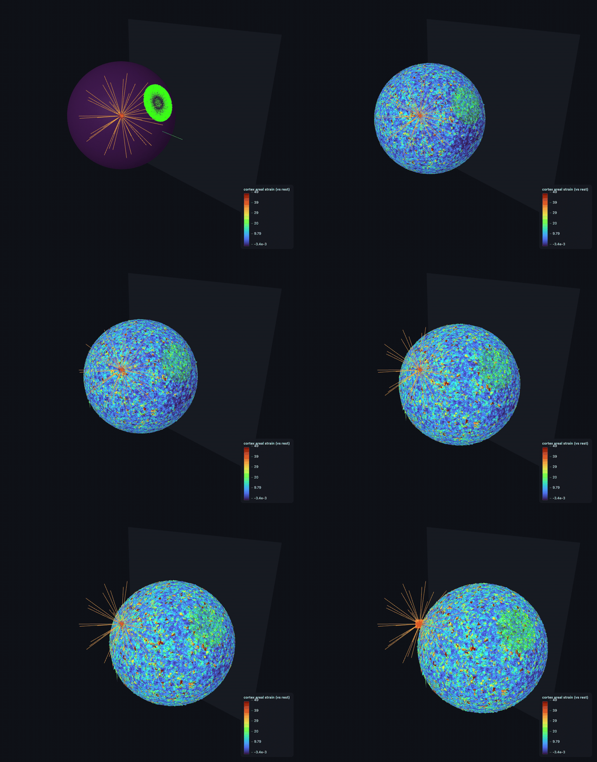
Native woven-actin cortex + microtubule aster + nucleus, with a green FA integrin-clutch patch gripping an explicit collagen-I Mikado fiber network — the substrate. As the cortex contracts the clutches pull the collagen, so the matrix visibly deforms: the cell recruits and remodels its own ECM, traction-driven and two-way (S6). The animation is the real viewer's own morph; the two stills are its von-Mises stress and areal-strain fields.
MCF7 cell recruiting a collagen-I matrix — shape
▶ shape — cortex + MT aster + collagen-I matrix (traction-driven remodel)
von-Mises stress morph
▶ σ_vm von-Mises stress (animated)
areal strain morph
▶ areal strain (animated)

⬇ Download full interactive viewer (304 MB)

FF — two-way collagen remodel: the cell densifies its own ECM (native, S6) ★ new

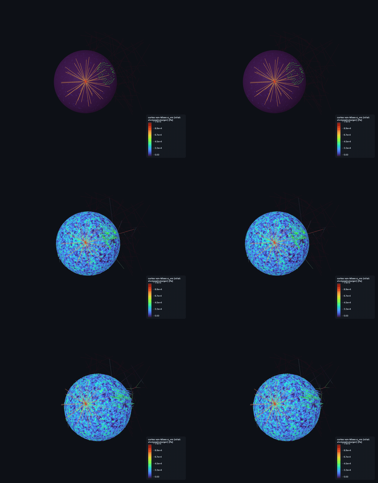
The native two-way ECM run. FA integrin clutches grip an explicit collagen-I Mikado matrix and contract it, so the matrix visibly densifies and reorients toward the cell — ~+265 nm densification, ~187 nN traction, per-clutch load at the physiological F*. Colour is collagen displacement (traction recruitment). The animation is the real viewer's own morph; the stills are its collagen-recruitment, von-Mises stress and areal-strain fields.
▶ shape — cortex + MT aster + FA patch gripping the collagen
▶ collagen recruitment / displacement (animated)
▶ σ_vm von-Mises stress (animated)
▶ areal strain (animated)

⬇ Download full interactive viewer (423 MB)

FF — substrate crawl: leading-edge protrusion + traction (native resolution)

The whole native-resolution cell crawling on a substrate: a polarized leading-edge cap protrudes, FA integrin clutches grip the surface, and the nucleus co-moves inside as the body translocates. The crawl is not scripted — it EMERGES from protrusion + clutch turnover, traction-driven at a physiological ~28–45 nm/s. The animation is the real viewer's own trajectory; the two stills are its von-Mises stress and areal-strain fields.
cell crawling on a substrate — shape
▶ shape — polarized protrusion + traction, nucleus co-moving
von-Mises stress morph
▶ σ_vm von-Mises stress (animated)
areal strain morph
▶ areal strain (animated)

⬇ Download full interactive viewer (209 MB)

FF — migration RESOLVED: the crawl engine driving on collagen (native resolution) ★ new

The headline migration result. The retrograde-flow crawl engine — which had never actually been wired to the ECM — is connected to an explicit collagen-I matrix with an F*-capped clutch, and the whole native cell now translocates by traction at a physiological, correctly poorly-motile MCF7 speed (~0.76 nm/s), pulling and reorienting the collagen as it moves. The animation is the real viewer's own morph; the two stills are its von-Mises stress and areal-strain fields.
▶ shape — the native cell crawling on collagen, traction-driven
▶ σ_vm von-Mises stress (animated)
▶ areal strain (animated)

⬇ Download full interactive viewer (264 MB)

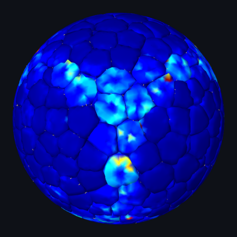
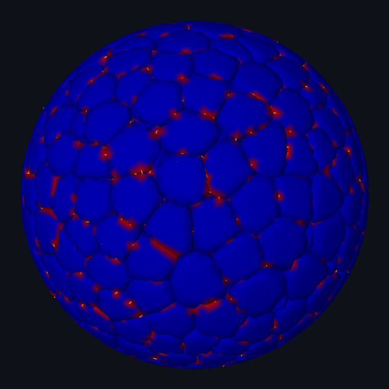
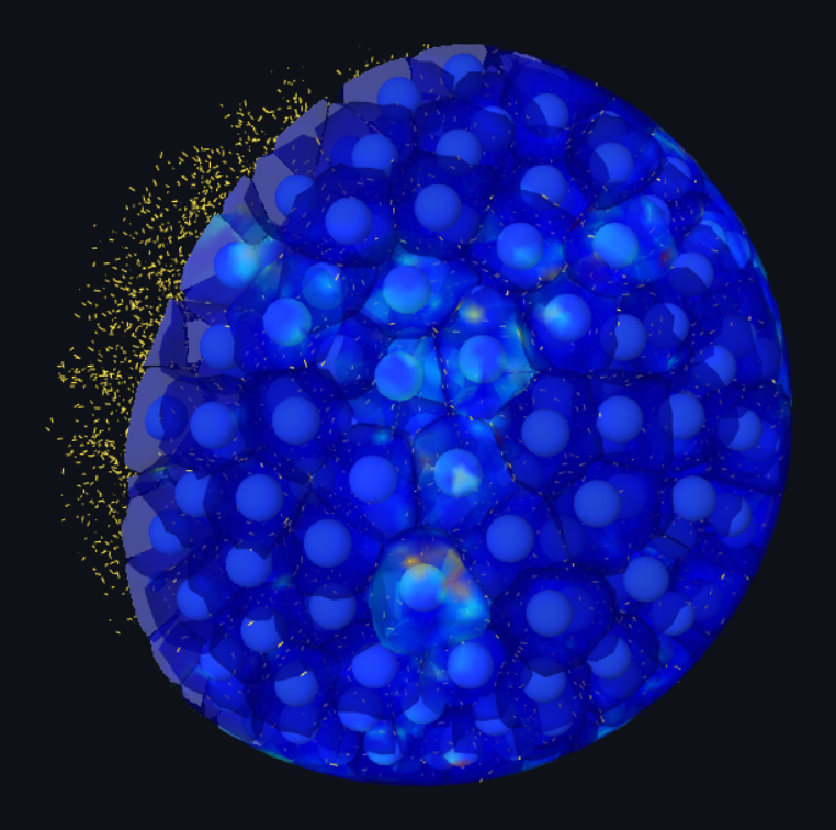
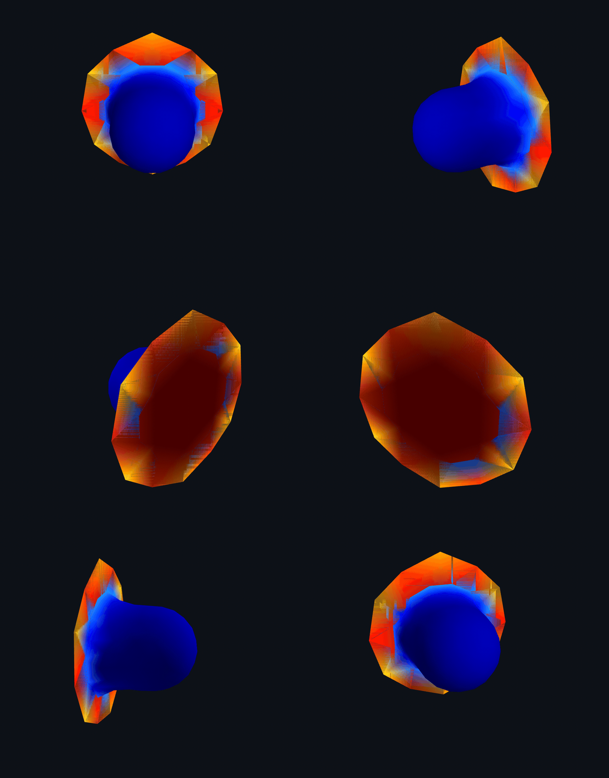
DCM — N=2,000 confluent spheroid, emergent stress + junctions

Per-cell virial von-Mises stress concentrating at load-bearing cadherin junctions; a rotating 3D turntable plus snapshots of the real viewer: surface stress, junction density, and a cut section exposing the per-cell nuclei.
N=2,000 confluent spheroid — 3D turntable of the faceted tissue
▶ 3D turntable — the faceted confluent spheroid rotating (von-Mises stress)
DCM N=2000 confluent spheroid — surface von-Mises stress + cadherin bonds
surface von-Mises stress + cadherin bonds
DCM N=2000 confluent spheroid — junction density
junction density
DCM N=2000 confluent spheroid — cut section — per-cell nuclei inside
cut section — per-cell nuclei inside

▶ Open interactive (42 MB) — rotate, recolour, cut, toggle nucleus

DCM ⊗ CBM hybrid — active spheroid assembly, then deformable refinement ★ new

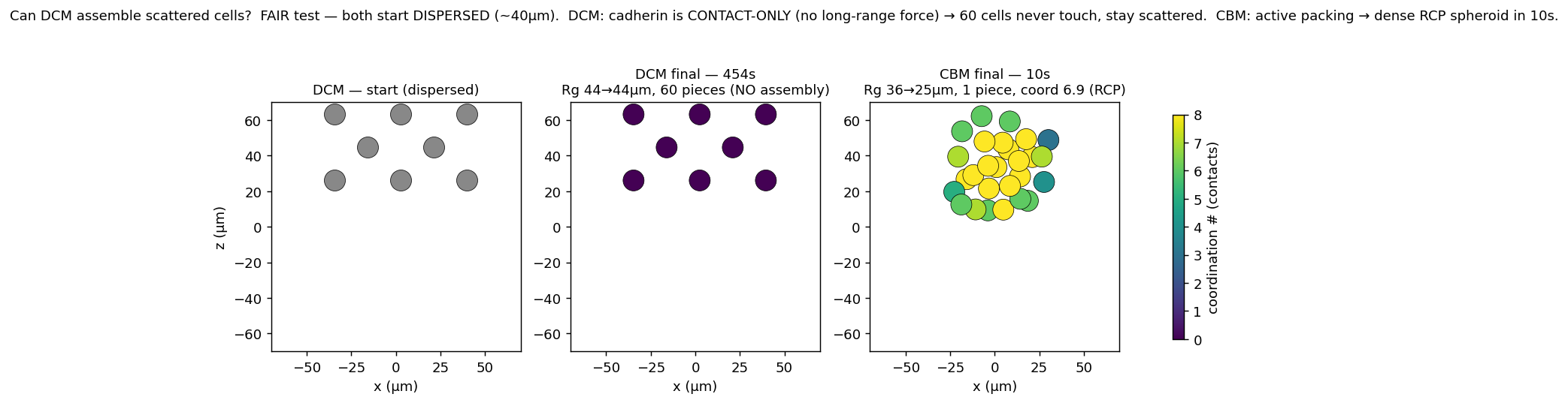
A new multi-scale route. DCM's cadherin is contact-only, so it cannot cheaply assemble dispersed cells — they never touch and stay scattered. A cheap center-based model (CBM: each cell a centre + radius + polarity under real forces, O(N)) packs hundreds of scattered cells into a dense random-close-packed spheroid in seconds — every force literature-anchored (Zbiral E_cell, MCF7 cadherin, Dessard viscosity), no magic number — then hands off to DCM for the deformable refinement. The full chain (assembly → force-driven rounding → refinement) runs at native N=400 in ~90 s; the two trajectories stitch into one seamless movie. The turntable is the assembled hybrid spheroid; open the live viewers to rotate the handoff init and the dish-assembly run.
▶ turntable — the CBM-assembled dense spheroid, handed to DCM (N=400)
FAIR head-to-head — DCM stays scattered (60 pieces, 454 s); CBM packs to dense RCP in 10 s

Honest gap: the compaction timescale (hours–days, cadherin-remodel-limited) is the open problem — the hybrid reaches the END-STATE by force, not its rate.

DCM — biology-time tissue flow: rate-driven T1 rearrangement ★ new

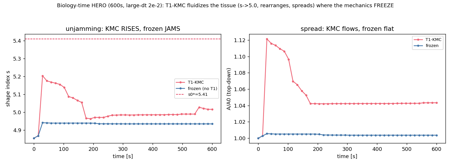
A confluent tissue jams and its mechanics freeze at large time-step (a genuine numerical fact, proven with a BDF2 A-stable integrator). The missing ingredient is not a better integrator but the physical rearrangement rate: a kinetic-Monte-Carlo T1 neighbour-exchange move at a physiological rate supplies the sub-Δt topological events the mechanics correctly skip, so the tissue fluidizes and flows over biology time. Over a 600 s run the T1-KMC tissue unjams (shape index s: 4.86 → 5.02, per-cell displacement 3.29 µm of real rearrangement, top-down A/A₀ → 1.04) while the frozen no-T1 control stays jammed (s 4.94, displacement 0.08 µm) — reaching the unjammed state ~20× faster in wall-clock (600 s of biology in ~40 min).
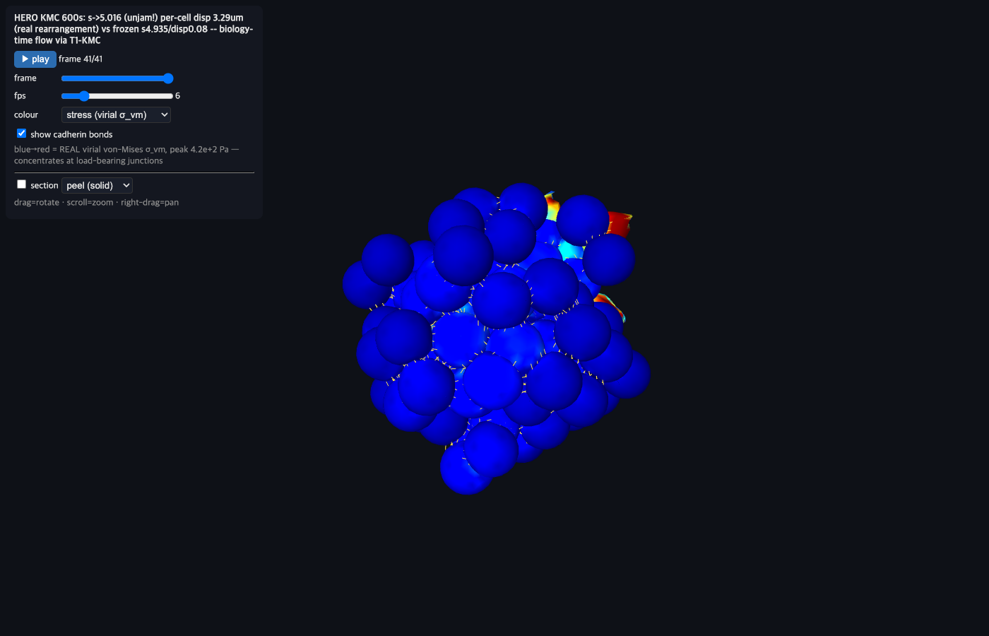
DCM T1-KMC 600s biology-time tissue, virial stress at junctions
the 600 s tissue (per-cell virial von-Mises stress, cadherin bonds) after T1-KMC fluidization — s→5.02, real rearrangement
shape-index and A/A0 trajectories: T1-KMC unjams and flows, frozen jams and stays flat
trajectories: T1-KMC (red) unjams + spreads vs the frozen no-T1 control (blue) that jams flat

Honest scope: the mechanics still freeze at this Δt — the rate-T1 supplies the flow, it does not remove the freeze; a transient early shape-drop and a modest final spread are open refinements. The point stands: emergent tissue creep is a fast sub-Δt T1 process, so a rate/event-driven move is the correct lever, not a stiffer solver.

DCM — active area-generation spreading (T-2) ★ new

The missing physics of single-cell spreading is active area generation: the cell does not merely stretch a fixed membrane, it actively grows its rest area A₀ (actin assembly at the basal interface), and the shell then flattens into a pancake against the substrate. This viewer plays that T-2 mechanism on a single deformable cell — drag to rotate, play the frames to watch it spread. Honest scope: this is a subdiv-2 (coarse) single-cell mechanism demonstration; at this resolution the periphery over-flares (a splay-inflation artifact), so it is shown as a mechanism proof-of-principle, with native-resolution confirmation still pending — never as a validated magnitude.
DCM active-area-generation spreading, cell flattening to a pancake
▶ the cell flattens to a spread pancake as its rest area A₀ actively grows (subdiv-2 demo)

▶ Open interactive (0.1 MB) — rotate & play the spreading

FF — 6-material ECM library: alignment, stiffness sensing & directional stress channeling ★ new

A grounded, alignment-controlled matrix library for the FF engine — collagen-I, fibrin, polyacrylamide, hyaluronic acid, Matrigel and agarose, each validated to its real modulus in SI Pascals (6/6 in-band). Two native-resolution viewers open live here: an aligned collagen matrix guiding cell contact, and a contractile inclusion whose stress channels down the fiber director. Drag to rotate; use the scene selector to sweep the nematic order S.
6 ECM materials in real Pascals
6/6 materials read out in real Pascals, each inside its literature band (collagen 12 Pa … agarose 14 kPa)
native biphasic stiffness sensing
Two-path biphasic stiffness sensing (native) — traction vs substrate modulus
directional stress decay exponent
Directional stress-decay exponent — aligned collagen channels stress well beyond isotropic

⬇ ECM library outputs & validation on GitHub

More interactive 3D viewers

Every cell is an explicit deformable body — drag to rotate, scroll to zoom.

▶ open 3D · 42 MB
N=2,000 confluent spheroid — stress + junctions (DCM)Per-cell von-Mises stress, cadherin-bond and nucleus overlays, live cut/peel. The flagship snapshotted above, fully interactive.
▶ open 3D · 14 MB
Native FF cell — membrane + cortex + nucleus + cytoplasmGPU-native woven-actin cortex with plasma membrane, nucleus and cytoplasm as explicit filaments (native resolution).
▶ open 3D
Voronoi spheroid + cadherin (N=400)Space-filling faceted cells held by explicit cadherin catch-bonds; membrane+cortex+cytoplasm+nucleus per cell.
▶ open 3D
Full-compartment spheroid (N=400)Confluent tissue: membrane + cortex + cytoplasm (turgor) + nucleus at MCF7 R_nuc=0.7R; IPC non-penetration.
▶ open 3D
Faceted confluent spheroid (N=400)Cells facet into space-filling polyhedra from differential cortical tension.
▶ open 3D
Cell division in a spheroid (N=400)Physically-anchored division; the spheroid grows cell-by-cell.
▶ open 3D
Emergent aggregation (N=100)100 cells self-assemble into a cohesive cluster via explicit cadherin catch-bonds.
▶ open 3D · 8 MB
Collagen-I recruitment over time (FF) Top-down: a native MCF7 cell's bound FA clutches grip an explicit collagen-I matrix and pull it as the cortex contracts — ▶ play the traction-driven displacement over ~100 s (fibers under the cell light up; clutches off → matrix frozen).
▶ open 3D
Filopodium into a collagen ECM (FF)Native-resolution actin bundle pushing into an explicit Mikado ECM network.
▶ open 3D
Filament architectures (FF)Cortex / filopodium / lamellipodium from one unified filament builder.

Validation against real data — shown honestly

Every mechanism is gated by a closed-form oracle (Hill, Bell–Evans, Storm–MacKintosh, Pereverzev, Young–Laplace, WLC) or an MCF7 / published experimental band. Below are the actual validation plots from 18 checkpoints across the ten weeks (plus the new 6-material ECM library and the migration-lever probes) — oracles pass tightly, and where the model is knowingly short of reality (the myosin γ-floor, the v1 spreading ceiling, the shear-modulus sourcing tension) the shortfall is annotated on the plot, not hidden.

v1 continuum top-down A/A0 spreading trajectory over 80 h, per ligand (Bare/Pre/Lam4)
△ short of reality — see the table below
DCM equilibrium A/A0 vs spheroid radius R fitted to the a+b/R+c/R^2 spreading law
△ short of reality — see the table below
Emergent motor-ensemble force-velocity v(F)/v0 (v0=1 um/s, F_stall=0.5 pN)
✓ passes oracle / analytic ground truth
Emergent Monte-Carlo bond off-rate k_off(F) biphasic catch-slip lifetime (min near F*=6.99 pN)
✓ passes oracle / analytic ground truth
Crosslinked-network strain-stiffening exponent beta in K(gamma) ~ gamma^beta
△ short of reality — see the table below
Point-force-dipole stress/displacement radial decay |sigma_xx(r)| in a semiflexible network
△ short of reality — see the table below
Single-fiber bending energy E_bend vs bead count n (FF Cytosim engine)
✓ passes oracle / analytic ground truth
DCM cell-cell doublet contact angle theta vs cadherin cohesion strength
△ short of reality — see the table below
DCM single-cell native volume conservation V/V0 under turgor over a BAOAB run
△ short of reality — see the table below
Gate-B emergent active cortical tension gamma on the connected ~38k-filament mesh (myoON vs myoOFF, rigid vs relaxed)
△ short of reality — see the table below
FF actomyosin cortical tension gamma_active vs myosin prestress f_myo (method-of-planes)
△ short of reality — see the table below
FF contractile network stress sigma vs per-link contractile force f_act and vs connectivity z
△ short of reality — see the table below
FF actin-network absolute shear modulus G vs crosslinker stiffness k_xl
△ short of reality — see the table below
FF whole-cell AFM apparent cortical tension gamma vs compressive strain (native N=70686, regulated dP=40 Pa)
△ short of reality — see the table below
FF biphasic-cytoplasm whole-cell Hertz apparent Young's modulus vs indentation regime
△ short of reality — see the table below
DCM (x) fiber-ECM cell-induced strain/stress radial decay slope (log-log)
△ short of reality — see the table below
FF single-cell actin-network structure/mechanics: mesh-size scaling, connectivity, floppy->rigid transition
△ short of reality — see the table below
FF biphasic-cytoplasm AFM validation — Hertz apparent modulus vs measured MCF7 (Zbiral 249 Pa): undrained 16.5x drains to ~5x
FF biphasic-cytoplasm AFM — Hertz apparent modulus vs measured MCF7 (Zbiral 249 Pa): the undrained 16.5× drains to ~4.7× as the cytoplasm is allowed to flow; the residual ~5× is the cortex, not the cytoplasm
△ short of reality — honest residual, annotated
ECM library consolidated validation dashboard
ECM library — consolidated native validation: 6/6 materials real-Pa, alignment→R_σ, stress-propagation n(S) vs KB-1.10
✓ all materials in literature band
fibrillar ECM strain stiffening
Fibrillar-ECM strain-stiffening — collagen/fibrin nonlinear modulus rising with applied strain
✓ passes oracle / analytic ground truth
FF migration regime lever probe
FF migration lever probe — the ECM regime (pore size / compliance) is the real ~2.3× lever; raw contractility & adhesion are not
△ short of reality — residual propulsion/solver limit, honest
The full validation table — 17 measured quantities vs. real data (oracle passes and honest shortfalls)
Measured quantityReal-world anchorHonest result vs. reality
v1 continuum top-down A/A0 spreading trajectory over 80 h, per ligand (Bare/Pre/Lam4)PI MCF7 spheroid experiment, Lam4/Pre/Bare (A/A0 = a + b/R + c/R^2), overlay-only
PI MCF7 experiment
Ligand ORDER (Bare<Pre<Lam4) and the a+b/R+c/R^2 functional form reproduced; platform raw at 82 h reaches A/A0 ~2.24-2.35 (Bare 2.24 / Pre 2.29 / Lam4 2.35) vs PI raw 9.3 (Bare) / 10.0 (Pre) / 13.5 (Lam4)
△ shortfall — Center-based continuum under-spreads ~4-6x (magnitude-gap decomposition x4.1/x4.4/x5.7; report headline states ~5-9x) below the PI values (overlay medians ~7.2-7.5 at 60 h; raw 9.3-13.5 at 82 h); a structural ceiling of the center-based 1-particle model, not a tuning gap (residual is cohesion-locked under-spread, core==hull so not a measurement artifact)
DCM equilibrium A/A0 vs spheroid radius R fitted to the a+b/R+c/R^2 spreading lawPI MCF7 spreading law (R=31-78 um: a=-0.33, b=188.7, c=-2655, r2=0.98), overlay-only
PI MCF7 experiment
Recovers the a+b/R+c/R^2 FORM; the r2=1.000 headline (h_dcm_gpu) is on only a 3-R mini-sweep with the RETRACTED basal-footprint metric (points ~= params, trivially high). Honest 5-point capstone fit is poor (r2=0.150, capstone_sweep.json) with c-sign OPPOSITE to PI (fit c=+2183 vs PI c=-2655, fit a=+4.76 vs PI -0.33); measured A/A0 ~1.6-2.5 (5 pts 1.62/2.48/1.73/1.58/2.48)
△ shortfall — A/A0 magnitude far below PI in the same R range and c-term sign disagrees; magnitude is a center/node structural limit. High-r2 fits are underdetermined (n~=3 points, 3 params); the footprint-normalised A/A0 is itself a PI-forbidden metric (top-down silhouette required)
Emergent motor-ensemble force-velocity v(F)/v0 (v0=1 um/s, F_stall=0.5 pN)Hill (1938) force-velocity closed form
closed-form oracle
Runtime tracks the Hill hyperbola exactly up to stall for three a/Fs ratios, then physically clamps v=0 beyond stall (the raw Hill form would go unphysically negative); D6 emergent-vs-oracle within +-5%
✓ matches within band
Emergent Monte-Carlo bond off-rate k_off(F) biphasic catch-slip lifetime (min near F*=6.99 pN)Pereverzev two-state catch-bond closed form (KU-2.5 / KU-2.18 params)
closed-form oracle
Emergent MC (n=20k per grid point) matches the closed-form oracle with max relative error 0.123%; PASSES the +-5% production gate across 1-60 pN (margin ~40x tighter than required)
✓ matches within band
Crosslinked-network strain-stiffening exponent beta in K(gamma) ~ gamma^betaStorm-MacKintosh semiflexible strain-stiffening regime (ratified yaml band |beta| in [1.5,2.5], 3/2 limit)
closed-form oracle
Ensemble slope |beta| = 1.530 (per-ramp 1.495/1.561/1.538), inside the [1.5,2.5] band
△ shortfall — Sits at the LOWER edge of the band (~3/2); the model does not reach the stronger-stiffening (~2) part of the Storm-MacKintosh regime
Point-force-dipole stress/displacement radial decay |sigma_xx(r)| in a semiflexible networkAnalytic force-dipole near-field 1/r^2 decay (brief band slope [-2.5,-1.5])
analytic ground truth
Ensemble (n=20 seeds) per-seed mean slope = -1.548 (fig annotated -1.549), inside the band and tracking the 1/r^2 reference (v7, after a HOOMD tag/row bond-virial bug fix retracted an earlier spurious -0.49 non-affine 'finding')
△ shortfall — Slope -1.55 lands at the SHALLOW edge (closer to 1/r^1.5 than 1/r^2); large per-seed scatter (std 0.906, thin grey lines span decades)
Single-fiber bending energy E_bend vs bead count n (FF Cytosim engine)Analytic worm-like-chain bending energy kappa*L/2R^2
analytic ground truth
FF corrected / analytic = 1.000 (<0.5% error, always-on); the raw discrete estimator under-counts by EXACTLY (n-2)/(n-1), and the end-correction recovers the analytic value
✓ matches within band
DCM cell-cell doublet contact angle theta vs cadherin cohesion strengthYoung-Dupre / Young-Laplace capillary balance (Maitre 2016 Nature doublet; SimuCell3D Supp Fig 5 oracle, cos theta = 1 - w_adh/gamma)
closed-form oracle
Measured theta rises 11deg -> 33deg with cohesion; at low adhesion measured 11deg vs Young-Dupre 13deg (delta 2deg) - cells DO flatten under adhesion per the analytic curve
△ shortfall — N=100 aggregate only PARTIALLY flattens (per-cell sphericity Psi 0.996 -> 0.972, regime-II); full tissue flattening is bond-density-limited (not reached)
DCM single-cell native volume conservation V/V0 under turgor over a BAOAB runAnalytic incompressibility V/V0 = 1 (volume-conserving turgor solver)
analytic ground truth
V/V0 holds near unity but drifts DOWN from ~0.95 to ~0.94 over 5000 steps (two-cell adhesion and 14-cell spheroid A/A0=2.36 sub-checks stay inside their stable bands)
△ shortfall — ~6% volume LEAK (V/V0 ~0.94, monotonically below 1) - the edge-spring-vs-turgor balance settles below exact conservation; exact setpoint needs turgor_inflate or k_edge recalibration
Gate-B emergent active cortical tension gamma on the connected ~38k-filament mesh (myoON vs myoOFF, rigid vs relaxed)MCF7 cortical-tension band IQR [0.18,0.40] mN/m, datum 0.27 (Hosseini 2020 Adv Sci, MCF-7 interphase)
published experiment
REFUTE: passive/turgor structural channel reaches only ~0.078-0.082 mN/m (rigid) and the MYOSIN-active g_soft channel ~1.4e-4 mN/m; gamma_active(rigid) = -0.0036 mN/m (adding myosin does not raise tension). Buckling shown NOT to be a lever
△ shortfall — Active-myosin cortical tension floored ~1000x under the MCF7 band; even the passive channel is ~3x under the 0.27 datum
FF actomyosin cortical tension gamma_active vs myosin prestress f_myo (method-of-planes)MCF7 IQR 0.18-0.40 mN/m and Salbreux band 0.35-0.65 mN/m; passive turgor gamma=dP*R/2
published experiment
At the literature NMIIA per-side stall (5 pN) gamma_active = 0.949 pN/um (~0.001 mN/m); even at f_myo=150 pN (super-physiological) only 28.5 pN/um (~0.03 mN/m). Passive turgor gamma=dP*R/2=665 pN/um reproduces the band
△ shortfall — Actomyosin-active tension floored ~100-1000x (native ~530x) under the MCF7/Salbreux band at the lit prestress; only the PASSIVE turgor channel reaches physiological tension
FF contractile network stress sigma vs per-link contractile force f_act and vs connectivity zSalbreux cortical-tension band 350-650 pN/um (0.35-0.65 mN/m)
published experiment
sigma stays UNDER the band across all f_act (sig_f 2.8->92 pN/um for f_act=1->100) AND flat across connectivity z=2.98-5.29 (sig_z 20-43 pN/um at f_act=20); at the NMIIA per-side stall 5 pN sigma ~10-15 pN/um (~25-50x under, doc headline ~30x)
△ shortfall — Confirms the active-gamma floor is robust: buckling and connectivity (z) do NOT lift it - the inextensible-filament regime cannot generate band-level active stress (even at super-physiological f_act=100 pN it is still ~4-7x under)
FF actin-network absolute shear modulus G vs crosslinker stiffness k_xlIn-vitro Kim/Gardel entropic band 0.07-1000 Pa and in-vivo cortex band ~0.1-1 kPa (analytic form G ~ f_na*k_xl*rho_L*lc)
published experiment
G is LINEAR in k_xl matching the analytic affine form (non-affine factor f_na=0.024 stable). With literature-sourced alpha-actinin k_xl=4.6e5 (Ferrer) G~8.6e5 Pa (~0.1-0.9 MPa regime); the broken default k_xl=0.1 gives 0.19 Pa (so k_xl=10 gives 19 Pa)
△ shortfall — With the SOURCED crosslinker stiffness the network shear modulus OVERSHOOTS the in-vivo cortex band (0.1-1 kPa) by ~10^2-10^3x; the in-band value (19 Pa) uses the ~4.5e6x-too-soft default knob - a genuine sourcing/units tension (PI-gated to correct live)
FF whole-cell AFM apparent cortical tension gamma vs compressive strain (native N=70686, regulated dP=40 Pa)MCF7 interphase cortical-tension band ~0.17 mN/m (Fischer-Friedrich)
published experiment
gamma_apparent ~0.150 mN/m over 0-60% strain, drifting up to 0.163 mN/m by ~78% strain (near the interphase datum); nucleus engages only past the contact strain (1 - R_nuc/R_cell = 0.667), as expected
△ shortfall — The apparent tension is turgor/cortex-PASSIVE (a dP=40 Pa Laplace proxy 0.5*dP*R_eq, not a force-fit of the indentation curve) and starts just BELOW the band; the active-myosin contribution remains floored ~0.0003 mN/m (see Gate-B / gamma-floor items)
FF biphasic-cytoplasm whole-cell Hertz apparent Young's modulus vs indentation regimeMeasured MCF7 Young's modulus, Zbiral 2023 E=249 Pa (10um colloidal, matched geometry)
published experiment
Undrained at Zbiral's fast rate 4114 Pa (16.5x); drained setpoint 40 Pa + K_drained 1548 Pa (6.2x); drained Lp + slow-ramp 1167 Pa (4.7x). Poroelastic drainage recovers most of the gap (tau_load ~0.1s << tau_osm ~30-200s)
△ shortfall — Whole-cell apparent modulus OVERSHOOTS measured MCF7 by ~5-16x; even fully drained a ~5x residual remains, attributed (rate-gated, open) to the over-stiff CORTEX lateral-bulge/crosslinker rather than the cytoplasm
DCM (x) fiber-ECM cell-induced strain/stress radial decay slope (log-log)Slater/Taeyoon-Kim fiber-ECM remodeling ~1/r decay in 2D (slope -1; d0sm01911a)
published experiment
Clean intermediate MID sub-band slope = -1.21 ~= -1, RECOVERING Kim ~1/r; contraction (cell -2%), focal-adhesion inward pull and radial alignment (|cos|~0.65) also reproduced
△ shortfall — 1/r holds only in the clean MID sub-band: near-field -0.63 (finite-cell, flat) and far-field +3.28 (finite-bed boundary) contaminate the whole-band slope, whose -0.19 is a fit-band artifact, not a real decay law
FF single-cell actin-network structure/mechanics: mesh-size scaling, connectivity, floppy->rigid transitionTaeyoon-Kim Brownian-dynamics actin-network model (MIT MS thesis, Kim 2007)
published experiment
Mesh xi ~ C_A^-0.48 matches the rho_L^-1/2 (C_A^-0.5) law; connectivity z=2R exactly; the elastic floppy->rigid rigidity-percolation transition (G rises steeply from ~0 sub-isostatic at z=0.2-0.6, through 0.18 at z=1, to ~7 pN/um^2 rigid at z=4) is reproduced (Head/MacKintosh/Kim)
△ shortfall — Match is structural/scaling only; the ABSOLUTE modulus inherits the crosslinker-stiffness overshoot-vs-floor problem (see kim_shear_modulus item) - shape/trend right, magnitude sourcing-dependent

Knowledge base — Contract-Graph, TAG & Obsidian

Every literature value, model decision and validation contract lives in a Notion Contract-Graph (8 atomic, bidirectionally-related databases) — the single source of truth. Two read layers are materialised from it: an Obsidian graph mirror and a DuckDB/TAG query engine. A controlled ablation measures how much each layer reduces LLM hallucination.

KB-architecture ablation (ffn_cellsim): as the KB evolved no-KB → RAG → +Obsidian graph → +TAG, only the TAG relational-query layer significantly improves structured KB-QA — and its win is total (accuracy 33% ≈ 29% ≈ 26% ≪ 100%; scaled study n=108, deterministic SQL gold, answerer claude-sonnet-4-6). RAG and the Obsidian graph alone add no statistically significant accuracy on structured questions. (TAG = Table-Augmented Generation, Biswal et al. 2024 — a NL→DuckDB-SQL query engine, NOT tags/labels.)

KB layer (condition)Accuracy (n=108)What it adds
C1 — no-KB (closed-book)33% micro / 53% macro [95% CI 24.1-42.6]Parametric memory only — nothing in context (the pre-KB ActiveCellSim era). Forced genuinely closed-book (--tools "", empty CWD) so it honestly refuses DB-specific questions instead of reading kb.duckdb; correctly rejects the 3 citation-trap fabrications and confirms the real Bell-1978 control.
C2 — RAG29% micro / 52% macro [95% CI 20.4-37.0]+ BM25 passages over the reference-PDF corpus (paper_chunks). Not significantly better than no-KB for structured QA (McNemar χ²=1.2, n.s.); has the worst citation grounding (16% unresolvable cites before/after the resolver fix).
C3 — RAG + Obsidian26% micro / 52% macro [95% CI 17.6-34.3]+ relevant Contract-Graph nodes and their typed neighbours (the graph the Obsidian mirror exposes). Best citation grounding (4% unresolvable) but still not significantly above no-KB for structured/relational/aggregation questions (McNemar χ²=0.4, n.s.).
C4 — RAG + TAG + Obsidian100% micro / 100% macro [95% CI 100-100]+ the synthesised DuckDB SQL result (exact join/aggregate) and the source_audit citation-integrity table. The only layer with a significant gain, and it is total — C4 ≥ every other condition on every one of the 108 questions; aggregation/relational/factual/lookup classes all switch on here.
Main scaled result (n=108): accuracy per condition with bootstrap 95% CI error bars (C1 33% / C2 29% / C3 26% overlap, C4 alone at 100%; McNemar C3→C4 80/80 wins, χ²=78, p≪.05) beside a per-class heatmap showing aggregation/relational/factual switch ON only under TAG.
RAGAS context-recall climbs 0.00→0.71 across C1→C4 — only TAG reliably retrieves the answer into context; faithfulness stays high everywhere.
KB data-flow schematic: one Notion Contract-Graph source-of-truth (8 atomic DBs) materialised into two read layers — the Obsidian ~640-node graph and kb.duckdb (8 tables + edges(808) + paper_chunks(5327) BM25) — both regenerated by refresh.sh and feeding the TAG engine.
The TAG loop (Biswal et al. 2024): syn (Claude turns the NL question into one DuckDB SELECT) → exec (read-only, SELECT-guarded, 1× self-repair on error) → gen (grounded answer + KB-id/DOI citations), fused with a BM25 paper_chunks semantic-excerpt branch.
Benchmark methodology & statistics (n=108 · deterministic SQL gold · McNemar · RAGAS / FActScore / TAG-Bench)

Scaled study: n=108 questions generated by gen_questions.py with gold computed by SQL over kb.duckdb (no LLM in the gold loop), stratified across aggregation(17)/relational(20)/lookup(49)/factual(14)/citation-trap(3)/citation-control(3)/negative(2). Bootstrap 95% CIs (kb_benchmark_stats.py): C1/C2/C3 overlap (~26-33%), C4 separate at 100%. McNemar paired tests on per-question correctness: C3 vs C4 — C4 wins 80, loses 0 → χ²=78.0, p≪.05; C1 vs C4 — wins 72, loses 0 → χ²=70.0, p≪.05; C1 vs C2 and C2 vs C3 NOT significant (χ²=1.2, 0.4). Study-1's apparent C1<C2<C3 staircase collapsed to C1≈C2≈C3≪C4 once n grew and CIs were added. syn is robust at scale: only 2/108 C4 SQL queries returned zero rows (0/86 on structured questions). Programmatic hallucination is low everywhere (3-6%) after a citation-resolver bug fix (rescore.py normalised bare-DOI vs https://doi.org/ forms; C4 unresolvable 20→5). Judge fixed at claude-opus-4-8, separate from the answerer.

Two tiers. Tier 1 — programmatic backbone (objective, no judge, all 108 questions): exact-match accuracy (gold substring/value present), hallucination rate (affirming a known-fabricated source, or citing a DOI/KB-id that does not resolve to a real kb.duckdb row), and citation groundedness (ALCE-style precision against the live DB). Tier 2 — standard LLM-evaluation metrics via a G-Eval-style judge (Liu 2023) on a stratified subset: RAGAS (Es 2023 — faithfulness, answer-relevancy, context-recall), FActScore/HaluEval (Min 2023; Li 2023 — decompose answer into atomic claims, score fraction unsupported = atomic hallucination rate), and TAG-Bench exact-match (Biswal 2024, the metric the TAG paper reports). RAGAS context-recall pins the causal mechanism: it climbs 0.00 → 0.04 → 0.28 → 0.96 across C1→C4 (scaled subset; 0.00→0.11→0.13→0.71 in the n=18 pilot) — only TAG reliably gets the answer INTO the context; relevancy (0.54→0.99) and EM (21→100%) follow. Faithfulness stays ~0.95-0.99 everywhere (answers grounded in whatever context given, incl. honest C1 refusals).

Obsidian graph — the Contract-Graph as a typed-link vault

The Obsidian mirror is the Contract-Graph's GRAPH read-layer — a read-only vault materialised FROM Notion (query_all → notion_to_obsidian.py; never hand-edited, fix Notion and refresh via refresh.sh). The 8 atomic Notion DBs (SourceEvidence, KnowledgeClaim, ModelContract, Parameter, ValidationGate, CodeMapping, RunResult, DecisionLedger) become a ~640-node typed-wikilink graph with bidirectional relations, augmented by Code/Test/Doc/DevLog nodes and supersedes/superseded_by supersession links. On disk it is a flat vault/ of ~1109 .md notes (one per Contract-Graph record plus per-paper reference stubs). Use it for visual graph exploration and 'what links to X' neighbourhood browsing; it cannot do joins/aggregation — those relational/aggregate questions go to the TAG/DuckDB query layer instead.

Obsidian Contract-Graph force-directed view: 1,050 connected notes coloured by node type (SourceEvidence, KnowledgeClaim, ModelContract, ValidationGate, Parameter, RunResult, CodeMapping, DevLog, Decision).

See the live data structure in Notion

The source-of-truth Contract-Graph itself — open the atomic databases directly (workspace access required):

SourceEvidence database ↗ Contract-Graph overview ↗ Knowledge-base entrypoint ↗

Engineering & optimization — how it runs fast enough to be native

Full-fidelity mechanistic physics is expensive; these are the optimization directions that make a native-resolution MCF7 cell (~266k particles) and N=2,000 spheroids tractable on a single workstation GPU — without ever coarse-graining the mechanism. Numbers are measured speedups, each validated by a bit-parity gate against the reference implementation.

GPU-native, differentiable runtime (NVIDIA Warp)

Both engines are GPU-resident and autodiff-differentiable on NVIDIA Warp — every force and integrator kernel runs on-device, enabling gradient-based parameter estimation / inverse design. HOOMD-blue is archived as the frozen parity oracle.

Custom BAOAB CUDA integrator — 44×

The Leimkuhler–Matthews BAOAB-limit constrained integrator was hand-written as a native C++/CUDA plugin — 44× faster than the per-step host path, and bit-parity identical to the reference (round-for-round wp.rint=np.round).

Linearly-implicit NF2007 solver — 10⁴×

The Nédélec–Foethke implicit step assembles the Jacobian analytically (bending 4th-difference + rank-1 crosslinks), LU-prefactored once or matrix-free CG on cupy — unconditionally stable, so dt is bounded by accuracy not stability: a 10⁴×-class speedup over explicit descent.

IPC contact absorbed into the solve

Incremental-Potential-Contact stiffness is fed straight into the matrix-free conjugate-gradient operator, so the implicit step absorbs contact — guaranteeing non-interpenetration (with CCD α-clamping) exactly where the old penalty contact tunnelled or diverged.

GPU-main: kill the per-step CPU sync

The binding updaters (myosin / crosslink / clutch KMC) and integrator were ported off per-step cpu_local_snapshot — which forced a GPU→CPU sync every step — onto gpu_local_snapshot / cupy and compiled CUDA kernels. Device-scalar CG reductions recover a further ~3–5× on the implicit step.

×40 mesoscale → then full native

The single sanctioned coarse-graining (×40 mesoscopic filament scale, PI-ratified as a hardware constraint) was the bridge; the going-forward path runs the full native cortex (--cortex-fil 38000, Nc≈266k) at ~5 s per simulated second on an RTX A5000 — the mechanism is never lumped, only the count is a ratified convention.

Parity-gating every kernel (~3×10⁻¹⁴)

Every ported kernel is gated bit-for-bit against the archived HOOMD-blue reference (FF all-Warp parity ~3×10⁻¹⁴) before it is trusted — optimization is only accepted when it does not change the physics, checked by a committed fixture test.

Fixed-size node pool + adaptive dt

A pre-allocated node pool with dormant slots keeps device topology static across division and remesh (no per-step reallocation; also fixes the HOOMD C++ rebuild memory leak), while a density-dependent drag η_eff(φ) raises the usable dt so the assembly/compaction timescale is reached in far fewer steps.

Design notes: ffn_sim/docs/v2_audit/ENGINE_ACCELERATION_PLAN_*.md, GPU_MAIN_PORT_*.md, PHASE_C_GPU_PARITY_*.md. Optimization is bounded by the mechanistic hard rule: an accelerated kernel must pass its parity gate, or it is rejected.